Images and videos
Images


Skull fractures
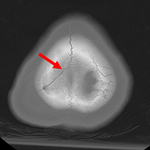
Gunshot wound with perpendicular blowout fracture
From the teaching collection of Demetrios Demetriades; used with permission
See this image in context in the following section/s:
Skull fractures
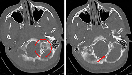
Fracture of temporal bone
From the teaching collection of Demetrios Demetriades; used with permission
See this image in context in the following section/s:
Skull fractures
Occipital fracture extending to foramen magnum: risk of brainstem compression by hematoma
From the teaching collection of Demetrios Demetriades; used with permission
See this image in context in the following section/s:
Skull fractures
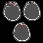
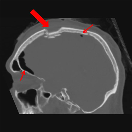
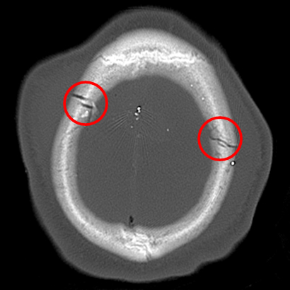
Sagittal CT images of an open, comminuted, depressed skull fracture. Note the associated pneumocephalus (small arrows). The level of depression is greater than the bony table and there are a number of bone fragments impacted below the inner cortex of the opposing bone (large arrow). Despite lack of underlying associated brain injury this fracture required operative debridement and elevation of the bone fragments. See also the corresponding coronal CT image
From the teaching collection of Demetrios Demetriades; used with permission
See this image in context in the following section/s:


Skull fractures
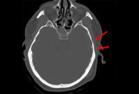
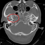
Fracture of temporal bone extending into foramen ovale
From the teaching collection of Demetrios Demetriades; used with permission
See this image in context in the following section/s:
Skull fractures
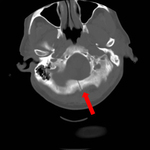
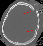
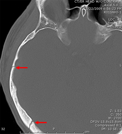
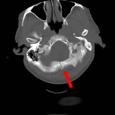
Axial CT scan showing nondepressed linear skull fracture (arrow) of the skull base involving the foramen magnum. This injury pattern is concerning for associated spinal fracture, cord injury, and blunt cerebrovascular injury
From the teaching collection of Demetrios Demetriades; used with permission
See this image in context in the following section/s:
Skull fractures
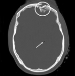
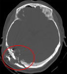
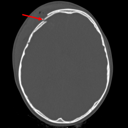
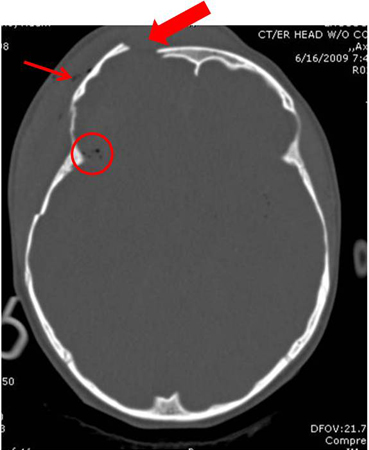
Axial CT scan demonstrating open elevated linear skull fracture (large arrow). Note the air in the soft tissues (small arrow), the small amount of pneumocephalus associated with the fracture (circle), and that the level of elevation of the bone fragment is significantly more than the thickness of the bony table
From the teaching collection of Demetrios Demetriades; used with permission
See this image in context in the following section/s:

Skull fractures
Gunshot wound with comminuted elevated fracture and pneumocephalus
From the teaching collection of Demetrios Demetriades; used with permission
See this image in context in the following section/s:


Skull fractures
Comminuted depressed fracture of the frontal sinus with air, fluid, and bone fragments in frontal sinus and pneumocephalus; level of depression greater than width of cortex
From the teaching collection of Demetrios Demetriades; used with permission
See this image in context in the following section/s:
Skull fractures
Transcranial gunshot wound
From the teaching collection of Demetrios Demetriades; used with permission
See this image in context in the following section/s:
Skull fractures
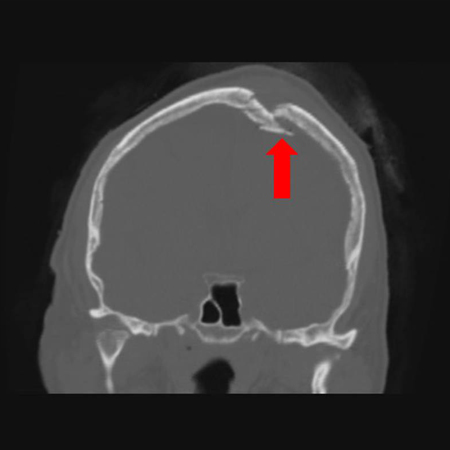
Coronal CT of an open, comminuted, depressed skull fracture. The level of depression is greater than the bony table and there are a number of bone fragments impacted below the inner cortex of the opposing bone (large arrow). Despite lack of underlying associated brain injury this fracture required operative debridement and elevation of the bone fragments. See also the corresponding sagittal CT image
From the teaching collection of Demetrios Demetriades; used with permission
See this image in context in the following section/s:
Use of this content is subject to our disclaimer