Ankle fractures
- Overview
- Theory
- Diagnosis
- Management
- Follow up
- Resources
Treatment algorithm
Please note that formulations/routes and doses may differ between drug names and brands, drug formularies, or locations. Treatment recommendations are specific to patient groups: see disclaimer
open fracture
emergency surgery
Open fractures require emergency treatment with a saline irrigation and debridement of the open wound and removal of all devitalized tissue as well as foreign debris by experienced surgeons.[49]British Orthopaedic Association and British Association of Plastic, Reconstructive and Aesthetic Surgeons. Open fractures. Dec 2017 [internet publication]. https://www.boa.ac.uk/standards-guidance/boasts.html The American Academy of Orthopaedic Surgeons (AAOS) recommends that patients with open fractures are brought to the operating room for debridement and irrigation as soon as possible, and ideally, within 24 hours postinjury.[39]American Academy of Orthopedic Surgeons. Prevention of surgical site infections after major extremity trauma: evidence-based clinical practice guideline. Mar 2022 [internet publication]. https://www.aaos.org/globalassets/quality-and-practice-resources/dod/ssitrauma/ssitraumacpg.pdf [40]American Academy of Orthopaedic Surgeons. Appropriate Use Criteria: prevention of surgical site infection after high energy extremity trauma. Mar 2022 [internet publication]. https://www.orthoguidelines.org/go/auc/auc.cfm?auc_id=225052 Treatment of the fracture is then with internal fixation when the wound is determined to be clean.[39]American Academy of Orthopedic Surgeons. Prevention of surgical site infections after major extremity trauma: evidence-based clinical practice guideline. Mar 2022 [internet publication]. https://www.aaos.org/globalassets/quality-and-practice-resources/dod/ssitrauma/ssitraumacpg.pdf [40]American Academy of Orthopaedic Surgeons. Appropriate Use Criteria: prevention of surgical site infection after high energy extremity trauma. Mar 2022 [internet publication]. https://www.orthoguidelines.org/go/auc/auc.cfm?auc_id=225052 In severely contaminated and comminuted fracture dislocations, temporizing external fixation may be required to facilitate repeated debridements, with delayed internal fixation.[39]American Academy of Orthopedic Surgeons. Prevention of surgical site infections after major extremity trauma: evidence-based clinical practice guideline. Mar 2022 [internet publication]. https://www.aaos.org/globalassets/quality-and-practice-resources/dod/ssitrauma/ssitraumacpg.pdf [40]American Academy of Orthopaedic Surgeons. Appropriate Use Criteria: prevention of surgical site infection after high energy extremity trauma. Mar 2022 [internet publication]. https://www.orthoguidelines.org/go/auc/auc.cfm?auc_id=225052 Emergency fracture management includes splinting of the affected limb.[19]Kyriacou H, Mostafa AMHAM, Davies BM, et al. Principles and guidelines in the management of ankle fractures in adults. J Perioper Pract. 2021 Nov;31(11):427-34. https://journals.sagepub.com/doi/full/10.1177/1750458920969029 http://www.ncbi.nlm.nih.gov/pubmed/33826430?tool=bestpractice.com
Antibiotics should be administered according to the type of open injury and severity of contamination at the time of diagnosis.[51]Gosselin RA, Roberts I, Gillespie WJ. Antibiotics for preventing infection in open limb fractures. Cochrane Database Syst Rev. 2004;(1):CD003764. http://www.ncbi.nlm.nih.gov/pubmed/14974035?tool=bestpractice.com Early delivery of antibiotics is suggested to lower the risk of deep infection in the setting of open fracture in major extremity trauma.[39]American Academy of Orthopedic Surgeons. Prevention of surgical site infections after major extremity trauma: evidence-based clinical practice guideline. Mar 2022 [internet publication]. https://www.aaos.org/globalassets/quality-and-practice-resources/dod/ssitrauma/ssitraumacpg.pdf [40]American Academy of Orthopaedic Surgeons. Appropriate Use Criteria: prevention of surgical site infection after high energy extremity trauma. Mar 2022 [internet publication]. https://www.orthoguidelines.org/go/auc/auc.cfm?auc_id=225052 [49]British Orthopaedic Association and British Association of Plastic, Reconstructive and Aesthetic Surgeons. Open fractures. Dec 2017 [internet publication]. https://www.boa.ac.uk/standards-guidance/boasts.html Utilization of preoperative antibiotics is suggested to prevent surgical site infections in operative treatment of open fractures.[39]American Academy of Orthopedic Surgeons. Prevention of surgical site infections after major extremity trauma: evidence-based clinical practice guideline. Mar 2022 [internet publication]. https://www.aaos.org/globalassets/quality-and-practice-resources/dod/ssitrauma/ssitraumacpg.pdf [40]American Academy of Orthopaedic Surgeons. Appropriate Use Criteria: prevention of surgical site infection after high energy extremity trauma. Mar 2022 [internet publication]. https://www.orthoguidelines.org/go/auc/auc.cfm?auc_id=225052 In patients with major extremity trauma undergoing surgery, the AAOS strongly recommends that antibiotic prophylaxis with systemic cefazolin or clindamycin be administered, except for type III (and possibly type II) open fractures, for which additional gram-negative coverage (e.g., piperacillin-tazobactam) is preferred.[39]American Academy of Orthopedic Surgeons. Prevention of surgical site infections after major extremity trauma: evidence-based clinical practice guideline. Mar 2022 [internet publication]. https://www.aaos.org/globalassets/quality-and-practice-resources/dod/ssitrauma/ssitraumacpg.pdf [40]American Academy of Orthopaedic Surgeons. Appropriate Use Criteria: prevention of surgical site infection after high energy extremity trauma. Mar 2022 [internet publication]. https://www.orthoguidelines.org/go/auc/auc.cfm?auc_id=225052 The AAOS also states that in patients with major extremity trauma undergoing surgery, local antibiotic prophylactic strategies, such as vancomycin powder, tobramycin-impregnated beads, or gentamicin-covered nails, may be beneficial.[39]American Academy of Orthopedic Surgeons. Prevention of surgical site infections after major extremity trauma: evidence-based clinical practice guideline. Mar 2022 [internet publication]. https://www.aaos.org/globalassets/quality-and-practice-resources/dod/ssitrauma/ssitraumacpg.pdf [40]American Academy of Orthopaedic Surgeons. Appropriate Use Criteria: prevention of surgical site infection after high energy extremity trauma. Mar 2022 [internet publication]. https://www.orthoguidelines.org/go/auc/auc.cfm?auc_id=225052 Recommended prophylactic regimens vary by region; therefore, local antibiotic protocols and advice from microbiology should be followed. Tetanus prophylaxis is administered depending on severity of injury and the tetanus status of the patient. Literature suggests that if tetanus immunization was more than 10 years prior, then administration of tetanus toxoid be included regardless of the wound pattern or type (i.e., tetanus-prone or not).[52]Rhee P, Nunley MK, Demetriades D, et al. Tetanus and trauma: a review and recommendations. J Trauma. 2005;58:1082-1088. http://www.ncbi.nlm.nih.gov/pubmed/15920431?tool=bestpractice.com The AAOS suggests wound coverage for fewer than 7 days from injury date.[39]American Academy of Orthopedic Surgeons. Prevention of surgical site infections after major extremity trauma: evidence-based clinical practice guideline. Mar 2022 [internet publication]. https://www.aaos.org/globalassets/quality-and-practice-resources/dod/ssitrauma/ssitraumacpg.pdf After closed fracture fixation, negative pressure wound therapy may mitigate the risk of revision surgery or the use of surgical site infection surveillance protocol. However, after open fracture fixation, negative pressure wound therapy does not appear to offer an advantage when compared with sealed dressings, as it does not decrease wound complications or amputations.[39]American Academy of Orthopedic Surgeons. Prevention of surgical site infections after major extremity trauma: evidence-based clinical practice guideline. Mar 2022 [internet publication]. https://www.aaos.org/globalassets/quality-and-practice-resources/dod/ssitrauma/ssitraumacpg.pdf Silver-coated dressings are not suggested to improve outcomes or decrease pin site infections.[39]American Academy of Orthopedic Surgeons. Prevention of surgical site infections after major extremity trauma: evidence-based clinical practice guideline. Mar 2022 [internet publication]. https://www.aaos.org/globalassets/quality-and-practice-resources/dod/ssitrauma/ssitraumacpg.pdf [40]American Academy of Orthopaedic Surgeons. Appropriate Use Criteria: prevention of surgical site infection after high energy extremity trauma. Mar 2022 [internet publication]. https://www.orthoguidelines.org/go/auc/auc.cfm?auc_id=225052
referral to vascular surgeon
Treatment recommended for ALL patients in selected patient group
Emergency treatment is required in those with vascular compromise, with urgent referral to a vascular surgeon or plastic surgeon for consideration of arterial damage. In cases where there is neurovascular compromise, anatomic realignment should be attempted.[50]Smit L, Boyle M. Management of ankle injuries in the prehospital environment - a review of the literature. Australas J Paramed. 2013;10:6. After reduction, the neurovascular status should be reassessed and documented. Adequate reduction should be confirmed by review of repeat radiographs.[7]British Orthopaedic Association Standards for Trauma. The management of ankle fractures. Aug 2016 [internet publication]. https://www.boa.ac.uk/static/f8b1c499-c38a-4805-8cb8d8eb3087bca7/8be763eb-5921-4cb2-b6802f3e65ce8e7f/the%20management%20of%20ankle%20fractures.pdf [43]Mordecai S, Al-Hadithy N. Management of ankle fractures. BMJ. 2011 Oct 28;343:d5204.
closed fracture + dislocation
closed reduction + splint
Emergency treatment is also required in those with a fracture dislocation.[23]Lawson KA, Ayala AE, Morin ML, et al. Republication of "ankle fracture-dislocations: a review". Foot Ankle Orthop. 2023 Aug 12;8(3):24730114231195058.
https://www.ncbi.nlm.nih.gov/pmc/articles/PMC10423454
http://www.ncbi.nlm.nih.gov/pubmed/37582190?tool=bestpractice.com
Radiographs should not be performed before reduction if they will cause an unacceptable delay.[7]British Orthopaedic Association Standards for Trauma. The management of ankle fractures. Aug 2016 [internet publication].
https://www.boa.ac.uk/static/f8b1c499-c38a-4805-8cb8d8eb3087bca7/8be763eb-5921-4cb2-b6802f3e65ce8e7f/the%20management%20of%20ankle%20fractures.pdf
Closed reduction is attempted with a short-leg splint or cast applied after successful reduction.[23]Lawson KA, Ayala AE, Morin ML, et al. Republication of "ankle fracture-dislocations: a review". Foot Ankle Orthop. 2023 Aug 12;8(3):24730114231195058.
https://www.ncbi.nlm.nih.gov/pmc/articles/PMC10423454
http://www.ncbi.nlm.nih.gov/pubmed/37582190?tool=bestpractice.com
The choice of short-leg splint or cast is based on fracture type, patient factors, surgical urgency, and clinician preference.[23]Lawson KA, Ayala AE, Morin ML, et al. Republication of "ankle fracture-dislocations: a review". Foot Ankle Orthop. 2023 Aug 12;8(3):24730114231195058.
https://www.ncbi.nlm.nih.gov/pmc/articles/PMC10423454
http://www.ncbi.nlm.nih.gov/pubmed/37582190?tool=bestpractice.com
[Figure caption and citation for the preceding image starts]: Backslab application: Webril is applied usually overlapping by about 50% to give at least 2 layers of padding under the plasterFrom the collection of B. Petrisor, MD; used with permission [Citation ends].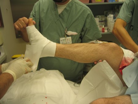 [Figure caption and citation for the preceding image starts]: Backslab application: the plaster slabs are then applied (2 side supports that wrap around the foot and one posterior support)From the collection of B. Petrisor, MD; used with permission [Citation ends].
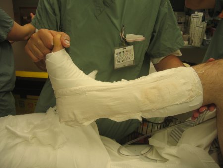 [Figure caption and citation for the preceding image starts]: Backslab application: an optional layer of Webril over the plaster prevents the tensor bandage or flannel from sticking and allows for ease of removal. The tensor bandage is then applied (some use flannel wrap) with minimal to no tension; it is "just rolled on"From the collection of B. Petrisor, MD; used with permission [Citation ends].
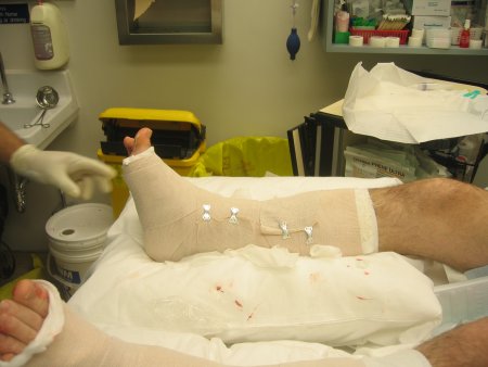
open reduction + fixation
Required if closed reduction is not successful.
referral to vascular surgeon
Treatment recommended for ALL patients in selected patient group
Emergency treatment is required in those with vascular compromise, with urgent referral to a vascular surgeon or plastic surgeon for consideration of arterial damage. In cases where there is neurovascular compromise, anatomic realignment should be attempted.[50]Smit L, Boyle M. Management of ankle injuries in the prehospital environment - a review of the literature. Australas J Paramed. 2013;10:6. After reduction, the neurovascular status should be reassessed and documented. Adequate reduction should be confirmed by review of repeat radiographs.[7]British Orthopaedic Association Standards for Trauma. The management of ankle fractures. Aug 2016 [internet publication]. https://www.boa.ac.uk/static/f8b1c499-c38a-4805-8cb8d8eb3087bca7/8be763eb-5921-4cb2-b6802f3e65ce8e7f/the%20management%20of%20ankle%20fractures.pdf [43]Mordecai S, Al-Hadithy N. Management of ankle fractures. BMJ. 2011 Oct 28;343:d5204.
isolated lateral malleolar fracture
short-leg cast
A short-leg cast can be given for 6 weeks, with weight-bearing in the cast for the last 3 weeks. In one randomized, multicentre, noninferiority trial, a 3-week period of immobilization proved noninferior to traditional 6 weeks of cast immobilisation for patients with stable, isolated Weber B type (transsyndesmotic) fibula fractures in their population, mean age 45 years.[53]Kortekangas T, Haapasalo H, Flinkkilä T, et al. Three week versus six week immobilisation for stable Weber B type ankle fractures: randomised, multicentre, non-inferiority clinical trial. BMJ. 2019 Jan 23;364:k5432. https://www.ncbi.nlm.nih.gov/pmc/articles/PMC6342249 http://www.ncbi.nlm.nih.gov/pubmed/30674451?tool=bestpractice.com
short-leg cast or internal fixation
A short-leg nonweight-bearing cast can be given for 6 weeks.
Operative fixation techniques for ankle fractures are varied. There are no guidelines for fixation devices.
Noncomminuted lateral malleolar fractures may be fixed with: 1) an interfragmentary lag screw and a neutralization plate; 2) posterior antiglide plate; 3) fibular intramedullary device.[19]Kyriacou H, Mostafa AMHAM, Davies BM, et al. Principles and guidelines in the management of ankle fractures in adults. J Perioper Pract. 2021 Nov;31(11):427-34. https://journals.sagepub.com/doi/full/10.1177/1750458920969029 http://www.ncbi.nlm.nih.gov/pubmed/33826430?tool=bestpractice.com
If the lateral malleolar fracture is comminuted, a bridge plate technique or intramedullary fixation may be used.[35]Lampridis V, Gougoulias N, Sakellariou A. Stability in ankle fractures: Diagnosis and treatment. EFORT Open Rev. 2018 May;3(5):294-303. https://www.ncbi.nlm.nih.gov/pmc/articles/PMC5994620 http://www.ncbi.nlm.nih.gov/pubmed/29951269?tool=bestpractice.com [56]Jain S, Haughton BA, Brew C. Intramedullary fixation of distal fibular fractures: a systematic review of clinical and functional outcomes. J Orthop Traumatol. 2014 Dec;15(4):245-54. https://link.springer.com/article/10.1007/s10195-014-0320-0 http://www.ncbi.nlm.nih.gov/pubmed/25304004?tool=bestpractice.com
After fixation of malleolar fractures, the syndesmosis should be tested for stability and, if unstable, fixation of the posterior malleolus or an anatomic reduction and stable fixation with syndesmotic screws are recommended.[35]Lampridis V, Gougoulias N, Sakellariou A. Stability in ankle fractures: Diagnosis and treatment. EFORT Open Rev. 2018 May;3(5):294-303. https://www.ncbi.nlm.nih.gov/pmc/articles/PMC5994620 http://www.ncbi.nlm.nih.gov/pubmed/29951269?tool=bestpractice.com
short-leg cast or internal fixation
A short-leg nonweight-bearing cast can be given for 6 weeks.[54]AO Surgery Reference. Infrasyndesmotic, medial fracture with lateral fracture/avulsion. 2024 [internet publication]. https://surgeryreference.aofoundation.org/orthopedic-trauma/adult-trauma/malleoli/infrasyndesmotic-medial-fracture-with-lateral-fracture-avulsion
Operative fixation techniques for ankle fractures are varied. There are no guidelines for fixation devices.
Noncomminuted lateral malleolar fractures may be fixed with: 1) an interfragmentary lag screw and a neutralization plate; 2) posterior antiglide plate; 3) fibular intramedullary device.[19]Kyriacou H, Mostafa AMHAM, Davies BM, et al. Principles and guidelines in the management of ankle fractures in adults. J Perioper Pract. 2021 Nov;31(11):427-34. https://journals.sagepub.com/doi/full/10.1177/1750458920969029 http://www.ncbi.nlm.nih.gov/pubmed/33826430?tool=bestpractice.com
If the lateral malleolar fracture is comminuted, a bridge plate technique or intramedullary fixation may be used.[35]Lampridis V, Gougoulias N, Sakellariou A. Stability in ankle fractures: Diagnosis and treatment. EFORT Open Rev. 2018 May;3(5):294-303. https://www.ncbi.nlm.nih.gov/pmc/articles/PMC5994620 http://www.ncbi.nlm.nih.gov/pubmed/29951269?tool=bestpractice.com [56]Jain S, Haughton BA, Brew C. Intramedullary fixation of distal fibular fractures: a systematic review of clinical and functional outcomes. J Orthop Traumatol. 2014 Dec;15(4):245-54. https://link.springer.com/article/10.1007/s10195-014-0320-0 http://www.ncbi.nlm.nih.gov/pubmed/25304004?tool=bestpractice.com
After fixation of malleolar fractures, the syndesmosis should be tested for stability and, if unstable, fixation of the posterior malleolus or an anatomic reduction and stable fixation with syndesmotic screws are recommended.[35]Lampridis V, Gougoulias N, Sakellariou A. Stability in ankle fractures: Diagnosis and treatment. EFORT Open Rev. 2018 May;3(5):294-303. https://www.ncbi.nlm.nih.gov/pmc/articles/PMC5994620 http://www.ncbi.nlm.nih.gov/pubmed/29951269?tool=bestpractice.com
internal fixation
Operative fixation techniques for ankle fractures are varied. There are no guidelines for fixation devices.
Noncomminuted lateral malleolar fractures may be fixed with: 1) an interfragmentary lag screw and a neutralization plate; 2) posterior antiglide plate; 3) fibular intramedullary device.
If the lateral malleolar fracture is comminuted, a bridge plate technique or intramedullary fixation may be used.[35]Lampridis V, Gougoulias N, Sakellariou A. Stability in ankle fractures: Diagnosis and treatment. EFORT Open Rev. 2018 May;3(5):294-303. https://www.ncbi.nlm.nih.gov/pmc/articles/PMC5994620 http://www.ncbi.nlm.nih.gov/pubmed/29951269?tool=bestpractice.com [56]Jain S, Haughton BA, Brew C. Intramedullary fixation of distal fibular fractures: a systematic review of clinical and functional outcomes. J Orthop Traumatol. 2014 Dec;15(4):245-54. https://link.springer.com/article/10.1007/s10195-014-0320-0 http://www.ncbi.nlm.nih.gov/pubmed/25304004?tool=bestpractice.com
After fixation of malleolar fractures, the syndesmosis should be tested for stability and, if unstable, fixation of the posterior malleolus or an anatomic reduction and stable fixation with syndesmotic screws are recommended.[35]Lampridis V, Gougoulias N, Sakellariou A. Stability in ankle fractures: Diagnosis and treatment. EFORT Open Rev. 2018 May;3(5):294-303. https://www.ncbi.nlm.nih.gov/pmc/articles/PMC5994620 http://www.ncbi.nlm.nih.gov/pubmed/29951269?tool=bestpractice.com
isolated medial malleolar fracture
short-leg cast
A short-leg nonweight-bearing cast can be given for 6 weeks.
internal fixation
Operative fixation techniques for ankle fractures are varied. There are no guidelines for fixation devices.
For medial malleolar fractures a buttress plate may be used for shearing fractures.[19]Kyriacou H, Mostafa AMHAM, Davies BM, et al. Principles and guidelines in the management of ankle fractures in adults. J Perioper Pract. 2021 Nov;31(11):427-34. https://journals.sagepub.com/doi/full/10.1177/1750458920969029 http://www.ncbi.nlm.nih.gov/pubmed/33826430?tool=bestpractice.com Transverse medial malleolar fractures may be fixed with 1 or 2 cancellous lag screws, tension band technique, or Kirschner wire fixation.[58]Ebraheim NA, Ludwig T, Weston JT, et al. Comparison of surgical techniques of 111 medial malleolar fractures classified by fracture geometry. Foot Ankle Int. 2014 May;35(5):471-7. http://www.ncbi.nlm.nih.gov/pubmed/24525543?tool=bestpractice.com
After fixation of malleolar fractures, the syndesmosis should be tested for stability and, if unstable, fixation of the posterior malleolus or an anatomic reduction and stable fixation with syndesmotic screws are recommended.[35]Lampridis V, Gougoulias N, Sakellariou A. Stability in ankle fractures: Diagnosis and treatment. EFORT Open Rev. 2018 May;3(5):294-303. https://www.ncbi.nlm.nih.gov/pmc/articles/PMC5994620 http://www.ncbi.nlm.nih.gov/pubmed/29951269?tool=bestpractice.com
bimalleolar/trimalleolar fracture
short-leg cast or internal fixation
A short-leg nonweight-bearing cast can be given for 6 weeks.
internal fixation
Operative fixation techniques for ankle fractures are varied. There are no guidelines for fixation devices.
Noncomminuted lateral malleolar fractures may be fixed with: 1) an interfragmentary lag screw and a neutralization plate; 2) posterior antiglide plate; 3) fibular intramedullary device.[19]Kyriacou H, Mostafa AMHAM, Davies BM, et al. Principles and guidelines in the management of ankle fractures in adults. J Perioper Pract. 2021 Nov;31(11):427-34. https://journals.sagepub.com/doi/full/10.1177/1750458920969029 http://www.ncbi.nlm.nih.gov/pubmed/33826430?tool=bestpractice.com
If the lateral malleolar fracture is comminuted, a bridge plate technique or intramedullary fixation may be used.[35]Lampridis V, Gougoulias N, Sakellariou A. Stability in ankle fractures: Diagnosis and treatment. EFORT Open Rev. 2018 May;3(5):294-303. https://www.ncbi.nlm.nih.gov/pmc/articles/PMC5994620 http://www.ncbi.nlm.nih.gov/pubmed/29951269?tool=bestpractice.com [56]Jain S, Haughton BA, Brew C. Intramedullary fixation of distal fibular fractures: a systematic review of clinical and functional outcomes. J Orthop Traumatol. 2014 Dec;15(4):245-54. https://link.springer.com/article/10.1007/s10195-014-0320-0 http://www.ncbi.nlm.nih.gov/pubmed/25304004?tool=bestpractice.com
For medial malleolar fractures a buttress plate may be used for shearing fractures.[19]Kyriacou H, Mostafa AMHAM, Davies BM, et al. Principles and guidelines in the management of ankle fractures in adults. J Perioper Pract. 2021 Nov;31(11):427-34. https://journals.sagepub.com/doi/full/10.1177/1750458920969029 http://www.ncbi.nlm.nih.gov/pubmed/33826430?tool=bestpractice.com Transverse medial malleolar fractures may be fixed with 1 or 2 cancellous lag screws, tension band technique, or Kirschner wire fixation.[58]Ebraheim NA, Ludwig T, Weston JT, et al. Comparison of surgical techniques of 111 medial malleolar fractures classified by fracture geometry. Foot Ankle Int. 2014 May;35(5):471-7. http://www.ncbi.nlm.nih.gov/pubmed/24525543?tool=bestpractice.com
Posterior malleolar fractures may be fixed with a posteriorly applied buttress plate or compression screws.[60]Tansey PJ, Chen J, Panchbhavi VK. Current concepts in ankle fractures. J Clin Orthop Trauma. 2023 Oct 16;45:102260. http://www.ncbi.nlm.nih.gov/pubmed/37872976?tool=bestpractice.com The decision to treat posterior malleolar fractures and the indications for treatment are controversial.[34]Stringfellow TD, Walters ST, Nash W, et al. Management of posterior malleolus fractures: a multicentre cohort study in the United Kingdom. Foot Ankle Surg. 2021 Aug;27(6):629-35. http://www.ncbi.nlm.nih.gov/pubmed/32878722?tool=bestpractice.com [60]Tansey PJ, Chen J, Panchbhavi VK. Current concepts in ankle fractures. J Clin Orthop Trauma. 2023 Oct 16;45:102260. http://www.ncbi.nlm.nih.gov/pubmed/37872976?tool=bestpractice.com Classic teaching suggests that fixation of the posterior malleolus be considered when the fracture size is greater than 25% to 33% of the joint surface. However, one meta-analysis suggests that this is based on low-quality evidence and that there is no consensus in the literature regarding the appropriate fracture size for fixation. The authors reported fixation of the medial and lateral malleoli is more important for overall stability.[64]van den Bekerom MP, Haverkamp D, Kloen P. Biomechanical and clinical evaluation of posterior malleolar fractures. A systematic review of the literature. J Trauma. 2009;66:279-84. http://www.ncbi.nlm.nih.gov/pubmed/19131839?tool=bestpractice.com There has been a move towards fixation of more posterior malleolar fractures, as even with small areas of the articular surface, there can be significant disruption to the posterior components of the syndesmosis.[60]Tansey PJ, Chen J, Panchbhavi VK. Current concepts in ankle fractures. J Clin Orthop Trauma. 2023 Oct 16;45:102260. http://www.ncbi.nlm.nih.gov/pubmed/37872976?tool=bestpractice.com [63]Odak S, Ahluwalia R, Unnikrishnan P, et al. Management of posterior malleolar fractures: a systematic review. J Foot Ankle Surg. 2016 Jan-Feb;55(1):140-5. http://www.ncbi.nlm.nih.gov/pubmed/26100091?tool=bestpractice.com The decision to treat posterior malleolar fractures may also be determined by other factors such as fracture displacement, congruency of the articular surface, and residual tibiotalar subluxation.[63]Odak S, Ahluwalia R, Unnikrishnan P, et al. Management of posterior malleolar fractures: a systematic review. J Foot Ankle Surg. 2016 Jan-Feb;55(1):140-5. http://www.ncbi.nlm.nih.gov/pubmed/26100091?tool=bestpractice.com Computed tomographic (CT) imaging may be helpful in defining fracture comminution, size, and configuration where fracture of the posterior malleolus is known or suspected.[7]British Orthopaedic Association Standards for Trauma. The management of ankle fractures. Aug 2016 [internet publication]. https://www.boa.ac.uk/static/f8b1c499-c38a-4805-8cb8d8eb3087bca7/8be763eb-5921-4cb2-b6802f3e65ce8e7f/the%20management%20of%20ankle%20fractures.pdf [33]Solan MC, Sakellariou A. Posterior malleolus fractures: worth fixing. Bone Joint J. 2017 Nov;99-B(11):1413-9. http://www.ncbi.nlm.nih.gov/pubmed/29092978?tool=bestpractice.com [34]Stringfellow TD, Walters ST, Nash W, et al. Management of posterior malleolus fractures: a multicentre cohort study in the United Kingdom. Foot Ankle Surg. 2021 Aug;27(6):629-35. http://www.ncbi.nlm.nih.gov/pubmed/32878722?tool=bestpractice.com
After fixation of malleolar fractures, the syndesmosis should be tested for stability and, if unstable, fixation of the posterior malleolus or an anatomic reduction and stable fixation with syndesmotic screws are recommended.[35]Lampridis V, Gougoulias N, Sakellariou A. Stability in ankle fractures: Diagnosis and treatment. EFORT Open Rev. 2018 May;3(5):294-303. https://www.ncbi.nlm.nih.gov/pmc/articles/PMC5994620 http://www.ncbi.nlm.nih.gov/pubmed/29951269?tool=bestpractice.com
If the fracture of the medial malleolus is minimally displaced after operative fixation of the fibula, nonoperative treatment yields similar results compared with operative treatment in terms of pain, development of osteoarthritis, and patient-reported functional outcome after a mean of 39 months.[59]Hoelsbrekken SE, Kaul-Jensen K, Mørch T, et al. Nonoperative treatment of the medial malleolus in bimalleolar and trimalleolar ankle fractures: a randomized controlled trial. J Orthop Trauma. 2013;27:633-637.
http://www.ncbi.nlm.nih.gov/pubmed/23454858?tool=bestpractice.com
[Figure caption and citation for the preceding image starts]: Mortise view of a trimalleolar fracture after fixation. Note: 2 syndesmosis screws were also usedFrom the collection of B. Petrisor, MD; used with permission [Citation ends].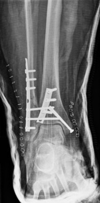 [Figure caption and citation for the preceding image starts]: Lateral view of a trimalleolar fracture after fixationFrom the collection of B. Petrisor, MD; used with permission [Citation ends].
 [Figure caption and citation for the preceding image starts]: Mortise view of a trimalleolar fracture dislocation with concomitant disruption of the syndesmosisFrom the collection of B. Petrisor, MD; used with permission [Citation ends].
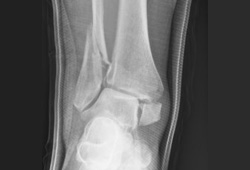
Choose a patient group to see our recommendations
Please note that formulations/routes and doses may differ between drug names and brands, drug formularies, or locations. Treatment recommendations are specific to patient groups. See disclaimer
Use of this content is subject to our disclaimer