Tests
1st tests to order
WBC count
erythrocyte sedimentation rate
Test
Usually elevated but may be normal; nonspecific, also elevated in other inflammatory conditions and in malignancy.[1][15] Can be used to monitor treatment; if persistently elevated after treatment, should trigger further assessment.[13] Often normal or only mildly elevated in chronic infection or in patients with a diabetic foot problem.[36][37] The trend in erythrocyte sedimentation rate may be useful for assessing improvement during treatment.[4][13]
Result
usually elevated
CRP
Test
Usually elevated. May be more helpful than the erythrocyte sedimentation rate because it normalizes more rapidly after successful treatment. Nonspecific.[4][13] Often normal or only mildly elevated in chronic infection or in patients with a diabetic foot problem.[36][37] The trend in CRP may be useful for assessing improvement during treatment.[4][13]
Result
usually elevated
plain x-rays of affected area
Test
Should be performed, as the initial imaging test, to look for evidence of osteomyelitis as well as for other relevant pathology such as fractures or bone tumors.[4][68][Figure caption and citation for the preceding image starts]: Plain x-ray of the left femur showing a lytic lesion in the medullary canal along with a "fallen leaf" sign with intramedullary sequestrum noted in the cavityCourtesy of the Oxford Bone Infection Unit; used with permission [Citation ends].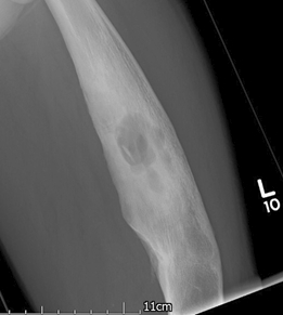
Plain films are an inherently insensitive test; radiologic changes may not be reliably present, especially in the early stages, and observer error may compound the issue, resulting in subtle changes being missed.[71] Plain radiographs do, however, provide an appropriate baseline examination for comparison as the disease progresses.
Subtle early radiographic findings of osteomyelitis include soft-tissue swelling and obscuration of the fat planes.[12][68] After 1-2 weeks, osteolysis, cortical loss, and periosteal reaction ensue.[68] Sequestra can sometimes be seen.
Result
subtle early findings include soft-tissue swelling and obscuration of fat planes, followed by osteolysis, cortical loss, periosteal reaction; sequestra can sometimes be seen; findings in chronic disease: intramedullary scalloping, cavities, and cloacae may be seen, with a "fallen leaf" sign noted when a piece of endosteal sequestrum has detached and fallen into the medullary canal
blood culture
Test
If blood cultures are indicated, samples should be taken before initiating antibiotics, whenever possible.[1][12] However, if a patient has suspected sepsis, antibiotic therapy should not be delayed.[1] In adults, blood cultures should be considered if the patient has a fever.[1] Blood cultures should be considered in all children.[12] The Infectious Diseases Society of America (IDSA) recommends obtaining bacterial (aerobic and anaerobic) blood cultures (2 sets) and baseline erythrocyte sedimentation rate and CRP in all patients with suspected native vertebral osteomyelitis.[13] Serologic tests for Brucella species should be obtained in patients with subacute native vertebral osteomyelitis residing in, or recently returning from, an area endemic for brucellosis.[13] Concomitant blood cultures for Brucella species are recommended but culture may be difficult and results slow to obtain, as the organism is intracellular and the number of circulating bacteria is usually low.[9][57] The laboratory should be notified when Brucella species is considered a potential cause of osteomyelitis so that cultures are examined only in a biologic safety cabinet.[9]
The IDSA further recommends that fungal blood cultures should be sought in patients with suspected native vertebral osteomyelitis who are at risk for fungal infection (epidemiologic risk or host risk factors).[13]
May facilitate early diagnosis, allowing treatment with antibiotics alone in children, in patients with acute infection, and in diabetic foot infections.
Result
may be positive, indicating the infecting organism and microbial sensitivities
MRI of bone
Test
Most helpful imaging modality because it gives good cross-sectional information about the bone and provides excellent evaluation of the adjacent soft tissues including abscess or fistulas.[12][68][72][73][74] MRI is also sensitive at depicting marrow signal changes of acute osteomyelitis.[68] MRI allows early detection of osteomyelitis; it is highly sensitive for detecting osteomyelitis as soon as 3-5 days after the onset of infection.[12][69][75] MRI spine is recommended in patients with suspected native vertebral osteomyelitis.[13] However, for all other types of suspected osteomyelitis, do not routinely use MRI as initial imaging. It is advised if additional imaging is required after initial radiographs have been taken.[68][76]
May show signs of infection in the medullary canal or surrounding soft tissues. Not useful in detecting cortical sequestra. Interpretation may require radiologists with a special interest in musculoskeletal imaging.
MRI is useful for the evaluation of osteomyelitis or soft tissue infection in the setting of extra-articular surgical hardware.[68] Advances in metal artifact reduction techniques have improved orthopedic hardware imaging, particularly in the appendicular skeleton.[68][77] However, MRI must be used with caution in the postoperative or post-trauma period because bone marrow and soft tissue edema may persist, therefore mimicking infection.[68][78]
In a child with acute hematogenous osteomyelitis who does not respond to medical therapy within 24-48 hours, or whose signs and symptoms suggest a potential role for surgical debridement, MRI may be performed to evaluate for an alternative diagnosis such as a malignancy.[4] However, do not order MRI in children until all appropriate clinical, laboratory and plain radiographic exams have been completed.[87]
The American College of Radiology recommends MRI for suspected osteomyelitis of the foot in patients with diabetes, after plain x-rays have been performed.[35] For more information, see Diabetic foot complications.
Result
may show high signal on T2 images or fat suppression sequences
guided bone biopsy or open bone biopsy
Test
Ideally, diagnosis should be confirmed by positive bacterial culture from deep microbiologic samples obtained via radiologic guided biopsy or open surgery.[4][9]
To maximize the sensitivity of microbiologic sampling it is advisable to stop antibiotics for at least 2 weeks before surgical debridement. However, in children with presumed acute hematogenous osteomyelitis who appear ill or have rapidly progressive infection, the Infectious Diseases Society of America (IDSA) recommends that empiric antibiotic treatment should be started immediately rather than withholding antibiotics until invasive procedures are performed.[4] In children with presumed acute hematogenous osteomyelitis who are not clinically ill and for whom an aspirate or biopsy by invasive diagnostic procedure is being planned prior to initiating antibiotics, the IDSA recommends that antibiotics should be withheld for no more than 48-72 hours.[4]
Bone biopsy is usually performed during the surgical debridement procedure.[1] Image-guided fine needle aspiration (FNA) is less disruptive to bone than biopsy and allows multiple samples to be taken. The IDSA recommends image-guided aspiration biopsy of a disc space or vertebral endplate in patients with suspected native vertebral osteomyelitis (based on clinical, laboratory, and imaging studies) when a microbiologic diagnosis for a known associated organism (Staphylococcus aureus, S lugdunensis, and Brucella species) has not been established by blood cultures or serologic tests.[13] Specimens should be submitted for Gram stain and aerobic and anaerobic culture and, if adequate tissue can be obtained, histopathology.[9] If results are negative or inconclusive (e.g., Corynebacterium species is isolated), a second imaging-guided aspiration biopsy, percutaneous endoscopic discectomy and drainage procedure, or open excisional biopsy, should be considered to collect additional specimens for repeat and additional testing.[9] In patients with neurologic compromise (with or without impending sepsis or hemodynamic instability), the IDSA recommends immediate surgical intervention and initiation of empiric antimicrobial therapy instead of withholding antimicrobial therapy prior to an image-guided diagnostic aspiration biopsy.[13] If adequate tissue can be safely obtained, specimens should be sent for pathologic examination to help confirm a diagnosis of native vertebral osteomyelitis and guide further diagnostic testing, especially in the setting of negative cultures.[13] Molecular diagnostics may be performed on bone biopsies but are not considered first-line diagnostic tests. Microorganism-specific nucleic acid amplification tests (NAATs) or a broader approach, such as 16S ribosomal RNA gene PCR/sequencing (for bacterial detection) may be considered.[9][62]
Result
may be positive, indicating the infecting organism and microbial sensitivities
Tests to consider
ultrasound
Test
May be a useful diagnostic tool when other modalities are not readily available.[68] Useful for regions that are complicated by orthopedic instrumentation and therefore might not be well seen with MRI.[69] Also helpful in guiding aspiration or biopsy for microbiologic diagnosis.[68]
Result
may show collections, subperiosteal abscesses, and adjacent joint infusions
CT scan
Test
Has a very limited role in diagnosis, but useful for visualizing extent of bone destruction; can identify small sequestra, cloacas, involucra, and intraosseous gas more reliably than MRI.[81][82][89] CT can help in the guidance of needle biopsies and joint aspiration; it is also valuable in cases of vertebral osteomyelitis.[69][80][81][82]
Do not order CT in children until all appropriate clinical, laboratory and plain radiographic exams have been completed.[87]
Result
bone destruction, sequestra, cloacas, involucra, and intraosseous gas
radionuclide scans
Test
Fluorodeoxyglucose positron emission tomography (FDG-PET) may be appropriate when initial radiographs are normal or show findings suggestive of osteomyelitis.[68] FDG-PET is helpful when it is difficult to determine whether an abnormality seen in the bone on MRI represents active infection or structural derangement of the bone. Recent fracture or orthopedic implant may lower accuracy of FDG-PET as FDG-uptake can be seen in inflammation, including aseptic hardware loosening.[68]
Result
increased uptake of radioactive injectate in infected sites
three-phase bone scans
Test
Three-phase bone scans use a radionuclide tracer, typically technetium-99-m (Tc99m) bound to a phosphorus-containing compound, that accumulates in areas of bone turnover and increased osteoblast activity.[79] Although there is insufficient evidence to recommend a three-phase bone scan for the initial evaluation of osteomyelitis, this modality may be appropriate when initial radiographs are normal or show findings suggestive of osteomyelitis.[68] A three-phase bone scan can be used to rule out osteomyelitis and has high sensitivity if conventional radiographs are normal and when bone is not affected by other underlying conditions such as osteoarthritis, recent fracture, or recent hardware implantation.[68][83] However, a positive three-phase bone scan is nonspecific, and other underlying bone abnormalities such as neuroarthropathy, trauma, surgery, or tumor reduce specificity markedly.[68][84][85] For patients with suspected native vertebral osteomyelitis, when MRI is not feasible (e.g., with implantable cardiac devices, cochlear implants, claustrophobia, or unavailability), a combination spine gallium/Tc99 bone scan, or CT scan, or a positron emission tomography scan can be considered.[13]
Result
increased uptake of radioactive injectate in infected sites
histology
Test
Deep sampling helps in the interpretation of microbiologic results. Some infections, such as tuberculosis and actinomycosis, can be directly diagnosed by histology alone.
In acute infection, direct microscopy with Gram staining of aspirated fluid gives a rapid indication of the type of organism present, but continued treatment should be based on full culture results with antibiotic sensitivities.[4][13] The presence of a sinus tract is pathognomonic of chronic osteomyelitis.[1][4][5][66]
In chronic osteomyelitis, Gram stain has a very low sensitivity and is of no practical use.
Histology can be used to confirm the diagnosis of culture-negative osteomyelitis by the demonstration of acute and chronic inflammatory cells, as well as dead bone, active bone resorption, and the presence of small sequestra.
In suspected cases of fracture-related osteomyelitis, the presence of ≥5 neutrophil polymorph counts per high-power field (x400 magnification) is a positive diagnostic test for infection.[67] In patients with mycetoma, a chronic soft tissue infection of the extremities which can extend into contiguous bone and connective tissue, sinus drainage may be examined grossly and microscopically for the presence of characteristic "sulfur granules".[9]
Result
may identify infecting organisms or inflammatory cells, necrotic bone adjacent to an inflammatory exudate, small sequestra
Use of this content is subject to our disclaimer