Approach
Both acute and chronic paronychia can be diagnosed based on their clinical characteristics; however, Gram stain, culture, and sensitivity are required for confirmation in the case of acute paronychia.
Acute paronychia
A clinical diagnosis of purulent infection in the nail folds should be confirmed with Gram stain, culture, and sensitivity, optimally after draining the infection under sterile conditions. The collection may already be draining spontaneously, in which case this fluid can be swabbed. MRSA should be suspected in all cases of acute paronychia; therefore, culture should be obtained in every case.
The patient with acute paronychia may be any age, although infants or toddlers (finger suckers) and adults with excessive water or contactant exposure are at highest risk. Women tend to be more commonly affected than men. Patients present complaining of pain in their nail folds, with or without a history of trauma or of drainage of pus.
Exam shows swollen proximal and/or lateral nail folds, often with a purulent collection in the skin.[Figure caption and citation for the preceding image starts]: Acute paronychiaFrom the collection of Dr N.J. Jellinek and Professor C.R. Daniel III [Citation ends]. This is tender and drains after incision with a number 11 blade, demonstrating frank pus.[Figure caption and citation for the preceding image starts]: Acute paronychia: incision with #11 bladeFrom the collection of Dr N.J. Jellinek and Professor C.R. Daniel III [Citation ends].
 [Figure caption and citation for the preceding image starts]: Acute paronychia: draining pusFrom the collection of Dr N.J. Jellinek and Professor C.R. Daniel III [Citation ends].
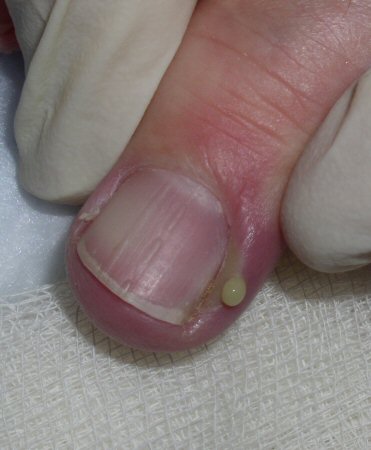 The diagnosis is confirmed through this clinical exam and the collection sent for Gram stain, culture, and sensitivity to aid in treatment.
Certain factors may point to herpes simplex infection as a cause for acute paronychia. These include lancinating pain, presence of vesicles, herpetiform arrangement of pustules and/or vesicles, or being refractory to antibacterial treatment. In these cases, a swab for Tzanck smear is indicated and, if negative, further tests for herpes simplex virus should be carried out.
With severe and/or refractory cases, an x-ray or magnetic resonance imaging (MRI) may be obtained to evaluate for underlying osteomyelitis.
Chronic paronychia
Diagnosis is clinical, based on typical inflamed nail folds with or without nail plate changes, with missing cuticle, in a person with risk factors for nail fold damage.
Patients are usually adults, and women more than men. Medication use should be noted as nail changes are well-documented among patients receiving certain chemotherapeutic agents (e.g., taxanes) and epidermal growth factor receptor inhibitors.[6][7] Other medications that can cause chronic paronychia include retinoids and protease inhibitors.[3] Nearly every patient will have a history of exposure to excessive water or contactants, including nail cosmetics (manicure, polish, gloss, cuticle treatment, hardeners). Patients complain of swelling and pain of the nail folds, occasionally with acute infectious episodes (in which case there may be a combination of acute and chronic features) and often irregular superficial nail plate abnormalities. On exam, there is swelling and shininess to the nail folds, a missing cuticle, and, frequently, superficial plate dystrophy.[Figure caption and citation for the preceding image starts]: Chronic paronychiaFrom the collection of Dr N.J. Jellinek and Professor C.R. Daniel III [Citation ends]. [Figure caption and citation for the preceding image starts]: Chronic paronychiaFrom the collection of Dr N.J. Jellinek and Professor C.R. Daniel III [Citation ends].
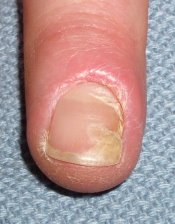
No laboratory tests are routinely ordered. A potassium hydroxide or fungal culture may show Candida; however, this colonization should not be confused with infection, and treatment of such is clearly secondary to irritant/moisture avoidance. A green nail may imply Pseudomonas infection. This diagnosis can be confirmed by sending off a nail clipping sample for Gram stain, culture, and sensitivity testing.
For refractory cases, an x-ray or magnetic resonance imaging may be obtained to rule out underlying osseous pathology, or biopsy to evaluate for squamous cell carcinoma. However, the diagnosis of chronic paronychia is usually made simply on clinical grounds.
Retronychia
Chronic paronychia and interruption of nail growth may rarely occur subsequent to posttraumatic ingrowth of the proximal nail plate into the proximal nail fold (retronychia) and can also occur after nail avulsion.[4][5] Important clinical criteria for diagnosis of retronychia include: inflammation of the proximal nail fold; granulation tissue emerging from under the nail fold; thickening of the proximal portion of the nail plate; and interruption of nail growth.[9]
Use of this content is subject to our disclaimer