Images and videos
Images

Graves disease
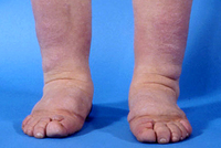
Pretibial myxedema (nonpitting edema)
Courtesy of Dr Vahab Fatourechi
See this image in context in the following section/s:
Graves disease
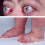
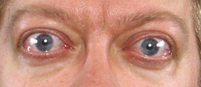
Lid retraction, mild proptosis, and mild chemosis
Courtesy of Dr Vahab Fatourechi
See this image in context in the following section/s:

Graves disease
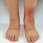
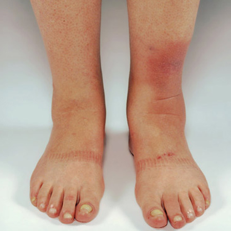
Elephantiasis
Courtesy of Dr Vahab Fatourechi
See this image in context in the following section/s:
Graves disease
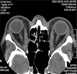
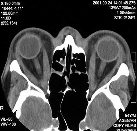
Axial CT scan through the orbits of a patient with Graves orbitopathy showing increased thickness of medial recti
Courtesy of Dr Petros Perros
See this image in context in the following section/s:

Graves disease
Orbitopathy and elephantiasis
Courtesy of Dr Vahab Fatourechi
See this image in context in the following section/s:

Graves disease
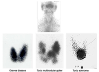
Iodine uptake scans. Typical appearances of absent uptake in thyroiditis (top panel). Diffuse increased uptake in Graves disease (lower left panel). Areas of increased and decreased uptake in toxic multinodular goiter (lower middle panel). Single area of increased uptake in a toxic adenoma (lower right panel)
Courtesy of Dr Petros Perros
See this image in context in the following section/s:
Use of this content is subject to our disclaimer