Acute cholangitis is an emergency that requires prompt diagnosis and treatment.[22]Buxbaum JL, Buitrago C, Lee A, et al. ASGE guideline on the management of cholangitis. Gastrointest Endosc. 2021 Aug;94(2):207-21.e14.
http://www.ncbi.nlm.nih.gov/pubmed/34023065?tool=bestpractice.com
Diagnosis is based on typical history and laboratory findings and confirmed by imaging, endoscopic retrograde cholangiopancreatography (ERCP), or endoscopic ultrasound-guided biliary drainage (EUS-BD) or percutaneous transhepatic biliary drainage if ERCP is unsuccessful.
Most patients will present with fever, jaundice, and right upper quadrant (RUQ) pain (Charcot triad), although some patients with a significant infection may have a surprisingly benign appearance overall.[3]Rumsey S, Winders J, MacCormick AD. Diagnostic accuracy of Charcot's triad: a systematic review. ANZ J Surg. 2017 Apr;87(4):232-8.
http://onlinelibrary.wiley.com/doi/10.1111/ans.13907/full
http://www.ncbi.nlm.nih.gov/pubmed/28213923?tool=bestpractice.com
Patients with acute cholangitis typically have diffuse RUQ pain and not classic Murphy sign.
Acute deterioration
Acute cholangitis can quickly become an acute, septic, life-threatening infection if not identified and treated promptly. Consider sepsis if there is acute deterioration in a patient in whom there is clinical evidence or strong suspicion of infection.[23]Singer M, Deutschman CS, Seymour CW, et al. The third international consensus definitions for sepsis and septic shock (Sepsis-3). JAMA. 2016 Feb 23;315(8):801-10.
https://jamanetwork.com/journals/jama/fullarticle/2492881
http://www.ncbi.nlm.nih.gov/pubmed/26903338?tool=bestpractice.com
The patient with sepsis may present with nonspecific or nonlocalized symptoms (e.g., acutely unwell with a normal temperature) or there may be severe signs with evidence of multiorgan dysfunction and shock.[23]Singer M, Deutschman CS, Seymour CW, et al. The third international consensus definitions for sepsis and septic shock (Sepsis-3). JAMA. 2016 Feb 23;315(8):801-10.
https://jamanetwork.com/journals/jama/fullarticle/2492881
http://www.ncbi.nlm.nih.gov/pubmed/26903338?tool=bestpractice.com
Sepsis represents the severe, life-threatening end of infection.
It is important to use systematic evaluation and recording of vital signs, alongside your clinical judgment, to assess the risk of deterioration due to sepsis.[23]Singer M, Deutschman CS, Seymour CW, et al. The third international consensus definitions for sepsis and septic shock (Sepsis-3). JAMA. 2016 Feb 23;315(8):801-10.
https://jamanetwork.com/journals/jama/fullarticle/2492881
http://www.ncbi.nlm.nih.gov/pubmed/26903338?tool=bestpractice.com
Treatment should be started immediately if a senior clinical decision-maker makes a diagnosis of suspected sepsis.[24]Evans L, Rhodes A, Alhazzani W, et al. Surviving sepsis campaign: international guidelines for management of sepsis and septic shock 2021. Crit Care Med. 2021 Nov 1;49(11):e1063-143.
https://journals.lww.com/ccmjournal/fulltext/2021/11000/surviving_sepsis_campaign__international.21.aspx
http://www.ncbi.nlm.nih.gov/pubmed/34605781?tool=bestpractice.com
Local protocols should be followed for the investigation and treatment of all patients with suspected sepsis, or those at risk of deterioration to sepsis.[24]Evans L, Rhodes A, Alhazzani W, et al. Surviving sepsis campaign: international guidelines for management of sepsis and septic shock 2021. Crit Care Med. 2021 Nov 1;49(11):e1063-143.
https://journals.lww.com/ccmjournal/fulltext/2021/11000/surviving_sepsis_campaign__international.21.aspx
http://www.ncbi.nlm.nih.gov/pubmed/34605781?tool=bestpractice.com
[25]UK Sepsis Trust. Sepsis: clinical tools. 2024 [internet publication].
https://sepsistrust.org/professional-resources/clinical-tools
[26]Daniels R, Nutbeam T, McNamara G, et al. The sepsis six and the severe sepsis resuscitation bundle: a prospective observational cohort study. Emerg Med J. 2011 Jun;28(6):507-12.
https://emj.bmj.com/content/28/6/507.long
http://www.ncbi.nlm.nih.gov/pubmed/21036796?tool=bestpractice.com
For more detail on when to suspect sepsis and on its management, see Sepsis in adults.
History and physical exam
Risk factors for acute cholangitis may be present, most notably:[11]Higuchi R, Takada T, Strasberg SM, et al; Tokyo Guideline Revision Committee. TG13 miscellaneous etiology of cholangitis and cholecystitis. J Hepatobiliary Pancreat Sci. 2013 Jan;20(1):97-105.
http://link.springer.com/article/10.1007%2Fs00534-012-0565-z/fulltext.html
http://www.ncbi.nlm.nih.gov/pubmed/23307005?tool=bestpractice.com
Older age (average age 50-60 years)[9]Ahmed M. Acute cholangitis - an update. World J Gastrointest Pathophysiol. 2018 Feb 15;9(1):1-7.
https://www.wjgnet.com/2150-5330/full/v9/i1/1.htm
http://www.ncbi.nlm.nih.gov/pubmed/29487761?tool=bestpractice.com
[16]Lavillegrand JR, Mercier-Des-Rochettes E, Baron E, et al. Acute cholangitis in intensive care units: clinical, biological, microbiological spectrum and risk factors for mortality: a multicenter study. Crit Care. 2021 Feb 6;25(1):49.
https://ccforum.biomedcentral.com/articles/10.1186/s13054-021-03480-1
http://www.ncbi.nlm.nih.gov/pubmed/33549136?tool=bestpractice.com
Known cholelithiasis
Underlying pancreaticobiliary disease (e.g., biliary strictures, primary or secondary sclerosing cholangitis)
Prior ERCP
Surgical or radiologic biliary tree intervention
HIV infection.
Most patients will present with Charcot triad (fever, jaundice, and RUQ pain).
Fever and RUQ pain are present in 65% to 90% of patients, though fever can be absent in older patients (those over 60 years old). About 65% of patients will have RUQ tenderness.[1]Kiriyama S, Kozaka K, Takada T, et al. Tokyo Guidelines 2018: diagnostic criteria and severity grading of acute cholangitis (with videos). J Hepatobiliary Pancreat Sci. 2018 Jan;25(1):17-30.
https://onlinelibrary.wiley.com/doi/full/10.1002/jhbp.512
http://www.ncbi.nlm.nih.gov/pubmed/29032610?tool=bestpractice.com
Jaundice is present in 60% to 70% of patients.[1]Kiriyama S, Kozaka K, Takada T, et al. Tokyo Guidelines 2018: diagnostic criteria and severity grading of acute cholangitis (with videos). J Hepatobiliary Pancreat Sci. 2018 Jan;25(1):17-30.
https://onlinelibrary.wiley.com/doi/full/10.1002/jhbp.512
http://www.ncbi.nlm.nih.gov/pubmed/29032610?tool=bestpractice.com
Other features may include:
Pale/putty/clay colored stools due to deficient bile secretion to the small intestine.
Pruritis (sensation of itch) associated with any liver disease.
Hypotension, which is present in 30% of patients.[1]Kiriyama S, Kozaka K, Takada T, et al. Tokyo Guidelines 2018: diagnostic criteria and severity grading of acute cholangitis (with videos). J Hepatobiliary Pancreat Sci. 2018 Jan;25(1):17-30.
https://onlinelibrary.wiley.com/doi/full/10.1002/jhbp.512
http://www.ncbi.nlm.nih.gov/pubmed/29032610?tool=bestpractice.com
Mental status changes, which occur in about 15% of patients. Hypotension and mental status changes are indicative of severe disease and are associated with a poor prognosis.[1]Kiriyama S, Kozaka K, Takada T, et al. Tokyo Guidelines 2018: diagnostic criteria and severity grading of acute cholangitis (with videos). J Hepatobiliary Pancreat Sci. 2018 Jan;25(1):17-30.
https://onlinelibrary.wiley.com/doi/full/10.1002/jhbp.512
http://www.ncbi.nlm.nih.gov/pubmed/29032610?tool=bestpractice.com
[27]Miura F, Okamoto K, Takada T, et al. Tokyo Guidelines 2018: initial management of acute biliary infection and flowchart for acute cholangitis. J Hepatobiliary Pancreat Sci. 2018 Jan;25(1):31-40.
https://onlinelibrary.wiley.com/doi/full/10.1002/jhbp.509
http://www.ncbi.nlm.nih.gov/pubmed/28941329?tool=bestpractice.com
When Charcot triad is associated with these features, it is referred to as Reynolds pentad.
Laboratory tests
Patients with suspected or known acute cholangitis should have the following tests done on admission (listed with common findings):
CBC: white blood cell count is typically >10,000/microliter (reference range 4800-10,800/microliter).
CRP (a marker of inflammation): can be elevated.
LFTs: hyperbilirubinemia is almost always present and, if absent, cholangitis is less likely to be the true diagnosis. Abnormal liver tests are usual findings, with elevated serum alkaline phosphatase and transaminases being typical.
BUN and creatinine: elevated renal function parameters are more common in severe disease states.
Electrolytes plus magnesium: possible decreases of serum potassium and magnesium.
Blood culture: positive blood culture rates among patients with acute cholangitis range from 21% to 71%.[28]Gomi H, Solomkin JS, Schlossberg D, et al. Tokyo Guidelines 2018: antimicrobial therapy for acute cholangitis and cholecystitis. J Hepatobiliary Pancreat Sci. 2018 Jan;25(1):3-16.
https://onlinelibrary.wiley.com/doi/full/10.1002/jhbp.518
http://www.ncbi.nlm.nih.gov/pubmed/29090866?tool=bestpractice.com
Bacteria are usually gram-negative, but gram-positive bacteria and anaerobes are also implicated in cholangitis.[28]Gomi H, Solomkin JS, Schlossberg D, et al. Tokyo Guidelines 2018: antimicrobial therapy for acute cholangitis and cholecystitis. J Hepatobiliary Pancreat Sci. 2018 Jan;25(1):3-16.
https://onlinelibrary.wiley.com/doi/full/10.1002/jhbp.518
http://www.ncbi.nlm.nih.gov/pubmed/29090866?tool=bestpractice.com
If sepsis is suspected, the following tests should also be ordered:
Coagulation profile: abnormalities can include decreased platelets and elevated prothrombin time.
Blood gas analysis and lactate: metabolic acidosis is common in severe disease states; raised lactate is associated with sepsis.
Transabdominal ultrasound and endoscopy
All patients presenting with RUQ pain and suspected cholangitis should undergo transabdominal ultrasound in the first instance.[30]Manes G, Paspatis G, Aabakken L, et al. Endoscopic management of common bile duct stones: European Society of Gastrointestinal Endoscopy (ESGE) guideline. Endoscopy. 2019 May;51(5):472-91.
https://www.thieme-connect.com/products/ejournals/abstract/10.1055/a-0862-0346
http://www.ncbi.nlm.nih.gov/pubmed/30943551?tool=bestpractice.com
[31]Bonomo RA, Edwards MS, Abrahamian FM, et al. 2024 clinical practice guideline update by the Infectious Diseases Society of America on complicated intraabdominal infections: diagnostic imaging of suspected acute cholecystitis and acute cholangitis in adults, children, and pregnant people. Clin Infect Dis. 2024 Oct 4;79(suppl 3):S104-8.
https://academic.oup.com/cid/article/79/Supplement_3/S104/7706134
http://www.ncbi.nlm.nih.gov/pubmed/38963820?tool=bestpractice.com
The main value of a transabdominal ultrasound is in detecting cholecystitis, which can mimic cholangitis, and to provide a limited evaluation of the biliary tree.
Transabdominal ultrasound is a quick, easy, and inexpensive initial diagnostic imaging modality. Its accuracy for detecting common bile duct (CBD) dilation is >90%, although the diameter of the CBD becomes a less useful parameter in patients who have previously undergone cholecystectomy (as physiologic dilation of the CBD can occur in this setting).
Transabdominal ultrasound has a poor sensitivity for detecting mid to distal CBD stones.[1]Kiriyama S, Kozaka K, Takada T, et al. Tokyo Guidelines 2018: diagnostic criteria and severity grading of acute cholangitis (with videos). J Hepatobiliary Pancreat Sci. 2018 Jan;25(1):17-30.
https://onlinelibrary.wiley.com/doi/full/10.1002/jhbp.512
http://www.ncbi.nlm.nih.gov/pubmed/29032610?tool=bestpractice.com
Patients with a history of biliary disease, an indwelling biliary prosthesis, or other predisposing factors should be considered for early ERCP for rapid diagnosis and therapy. [Figure caption and citation for the preceding image starts]: Endoscopic retrograde cholangiopancreatography reveals a large common bile duct (CBD) stone (arrow) in the mid-common bile ductFrom the collection of Douglas G. Adler; used with permission [Citation ends].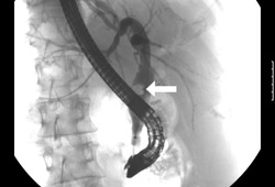 [Figure caption and citation for the preceding image starts]: Endoscopic photo of same patient following removal of large common bile duct (CBD) stone; note copious pus draining through the ampullaFrom the collection of Douglas G. Adler; used with permission [Citation ends].
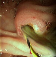
Subsequent imaging studies
If there is high clinical suspicion of cholangitis, and transabdominal ultrasound is negative, an abdominal computed tomography (CT) scan with intravenous contrast can be considered.[31]Bonomo RA, Edwards MS, Abrahamian FM, et al. 2024 clinical practice guideline update by the Infectious Diseases Society of America on complicated intraabdominal infections: diagnostic imaging of suspected acute cholecystitis and acute cholangitis in adults, children, and pregnant people. Clin Infect Dis. 2024 Oct 4;79(suppl 3):S104-8.
https://academic.oup.com/cid/article/79/Supplement_3/S104/7706134
http://www.ncbi.nlm.nih.gov/pubmed/38963820?tool=bestpractice.com
Abdominal CT provides better anatomical detail of the biliary tree than transabdominal ultrasound. It is particularly useful to visualize the distal part of the CBD.
If neoplasm is suspected as the cause of cholangitis, abdominal CT is a better initial imaging choice than transabdominal ultrasound.
CT scans are contraindicated in patients with intravenous contrast dye allergy and may be detrimental to those with renal dysfunction.
Magnetic resonance cholangiopancreatography (MRCP) is an excellent noninvasive magnetic resonance imaging (MRI) modality with good sensitivity and specificity for the diagnosis of biliary obstruction. It should be ordered if ultrasound and CT are negative and a high clinical suspicion remains for cholangitis.[30]Manes G, Paspatis G, Aabakken L, et al. Endoscopic management of common bile duct stones: European Society of Gastrointestinal Endoscopy (ESGE) guideline. Endoscopy. 2019 May;51(5):472-91.
https://www.thieme-connect.com/products/ejournals/abstract/10.1055/a-0862-0346
http://www.ncbi.nlm.nih.gov/pubmed/30943551?tool=bestpractice.com
[31]Bonomo RA, Edwards MS, Abrahamian FM, et al. 2024 clinical practice guideline update by the Infectious Diseases Society of America on complicated intraabdominal infections: diagnostic imaging of suspected acute cholecystitis and acute cholangitis in adults, children, and pregnant people. Clin Infect Dis. 2024 Oct 4;79(suppl 3):S104-8.
https://academic.oup.com/cid/article/79/Supplement_3/S104/7706134
http://www.ncbi.nlm.nih.gov/pubmed/38963820?tool=bestpractice.com
MRCP can provide cholangiograms and pancreatograms and can identify biliary stones, strictures, and/or pancreatic and biliary malignancies with a high sensitivity and specificity. It can also sometimes diagnose cholangitis based on the appearance of the bile duct walls.
MRCP can be particularly informative if there are confounding factors (e.g., underlying liver disease) that could lead to fever, RUQ pain, and jaundice.
MRI scans are contraindicated in some patients with metallic bioimplants.
While MRCP is diagnostically valuable, all patients with cholangitis will ultimately require biliary decompression, most commonly via ERCP. MRCP should thus not be viewed as a requisite study in patients with suspected cholangitis, but is often a helpful tool in determining an etiology and planning for definitive therapy.
Endoscopic ultrasound (EUS) is as accurate as MRCP for the detection of choledocholithiasis.[32]Verma D, Kapadia A, Eisen GM, et al. EUS vs MRCP for detection of choledocholithiasis. Gastrointest Endosc. 2006 Aug;64(2):248-54.
http://www.ncbi.nlm.nih.gov/pubmed/16860077?tool=bestpractice.com
It should be performed if there is suspicion of a CBD stricture or stone that has not been already seen on ultrasound or MRCP.[30]Manes G, Paspatis G, Aabakken L, et al. Endoscopic management of common bile duct stones: European Society of Gastrointestinal Endoscopy (ESGE) guideline. Endoscopy. 2019 May;51(5):472-91.
https://www.thieme-connect.com/products/ejournals/abstract/10.1055/a-0862-0346
http://www.ncbi.nlm.nih.gov/pubmed/30943551?tool=bestpractice.com
It can also be used if other imaging modalities are unsuitable for the patient.
If EUS is positive for CBD stones, ERCP can often be performed concomitantly. EUS can also facilitate biliary decompression via EUS-BD, if available.[33]Isayama H, Nakai Y, Itoi T, et al. Clinical practice guidelines for safe performance of endoscopic ultrasound/ultrasonography-guided biliary drainage: 2018. J Hepatobiliary Pancreat Sci. 2019 Jul;26(7):249-69.
https://onlinelibrary.wiley.com/doi/10.1002/jhbp.631
http://www.ncbi.nlm.nih.gov/pubmed/31025816?tool=bestpractice.com
[34]ASGE Standards of Practice Committee; Pawa S, Marya NB, Thiruvengadam NR, et al. American Society for Gastrointestinal Endoscopy guideline on the role of therapeutic EUS in the management of biliary tract disorders: summary and recommendations. Gastrointest Endosc. 2024 Dec;100(6):967-79.
https://www.giejournal.org/article/S0016-5107(24)00188-3/fulltext
http://www.ncbi.nlm.nih.gov/pubmed/39078360?tool=bestpractice.com
Surgical approaches
Surgery should be utilized for diagnostic purposes only when other modalities have failed to identify biliary stone(s) and/ or site(s) of obstruction, or when those modalities are not available, are not feasible, or are contraindicated. In practice, surgery is rarely required for diagnosis or treatment of cholangitis as less invasive approaches are almost always adequate.
