Images and videos
Images
Diffuse idiopathic skeletal hyperostosis
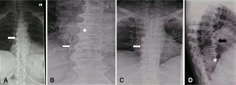
X-ray images of the thoracic spine of a patient with DISH. (A–C) Posterior–anterior and (D) lateral: large right-sided flowing bridges (white arrows). Note the space between the ligament and the vertebral body (*). Thick flowing ossification of the anterior lateral ligament is shown (black arrow)
Mader R, et al. RMD Open 2020; 6: e001151. doi: 10.1136; used with permission
See this image in context in the following section/s:
Diffuse idiopathic skeletal hyperostosis
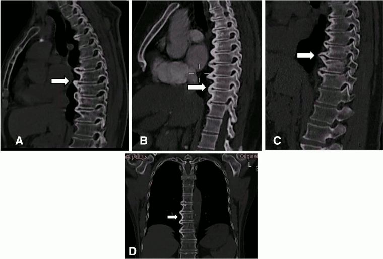
CT images of the thoracic spine in DISH. (A–C) Sagittal: CT scan images of anterior flowing osteophytes (arrows). (D) Coronal: dish of the thoracic spine (arrow) reconstructed from the chest CT scan. L = left
Mader R, et al. RMD Open 2020; 6: e001151. doi: 10.1136; used with permission
See this image in context in the following section/s:
Videos
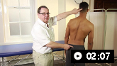 Inspection of the back
How to perform an inspection examination of the back, including inspection of gait and posture
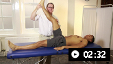 Physical examination of the back demonstration
A primary care physician demonstrates how to perform a physical examination of the back
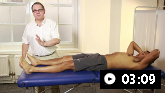 Neurological examination of the back demonstration
A primary care physician shows how to perform a neurological examination of the back
Use of this content is subject to our disclaimer