Sepsis is a spectrum of disease, where there is a systemic and dysregulated host response to an infection.[1]Singer M, Deutschman CS, Seymour CW, et al. The third international consensus definitions for sepsis and septic shock (Sepsis-3). JAMA. 2016 Feb 23;315(8):801-10.
https://jamanetwork.com/journals/jama/fullarticle/2492881
http://www.ncbi.nlm.nih.gov/pubmed/26903338?tool=bestpractice.com
Presentation may range from nonspecific or nonlocalized symptoms (e.g., feeling ill with a normal temperature), to severe signs with evidence of multiorgan dysfunction and septic shock. Risk of progression to fulminant disease is determined by various factors, including:
Magnitude and nature of the infective focus;
Timeliness and quality of interventions; and
Genetic and acquired predisposition of the patient.
The importance of early recognition of suspected sepsis
Early recognition of sepsis is essential because early treatment - when sepsis is suspected but is yet to be confirmed - is associated with significant short- and long-term benefits in outcome.[10]National Institute for Health and Care Excellence. Suspected sepsis: recognition, diagnosis and early management. March 2024 [internet publication].
https://www.nice.org.uk/guidance/ng51
[65]Rivers E, Nguyen B, Havstad S, et al. Early goal-directed therapy in the treatment of severe sepsis and septic shock. N Engl J Med. 2001 Nov 8;345(19):1368-77.
https://www.nejm.org/doi/10.1056/NEJMoa010307
http://www.ncbi.nlm.nih.gov/pubmed/11794169?tool=bestpractice.com
[66]Gao F, Melody T, Daniels R, et al. The impact of compliance with 6-hour and 24-hour sepsis bundles on hospital mortality in patients with severe sepsis: a prospective observational study. Crit Care. 2005;9(6):R764-70.
https://ccforum.biomedcentral.com/articles/10.1186/cc3909
http://www.ncbi.nlm.nih.gov/pubmed/16356225?tool=bestpractice.com
[67]Jones AE, Focht A, Horton JM, et al. Prospective external validation of the clinical effectiveness of an emergency department-based early goal-directed therapy protocol for severe sepsis and septic shock. Chest. 2007 Aug;132(2):425-32.
https://www.ncbi.nlm.nih.gov/pmc/articles/PMC2703721
http://www.ncbi.nlm.nih.gov/pubmed/17573521?tool=bestpractice.com
[68]Puskarich MA, Marchick MR, Kline JA, et al. One year mortality of patients treated with an emergency department based early goal directed therapy protocol for severe sepsis and septic shock: a before and after study. Crit Care. 2009;13(5):R167.
https://ccforum.biomedcentral.com/articles/10.1186/cc8138
http://www.ncbi.nlm.nih.gov/pubmed/19845956?tool=bestpractice.com
[69]Daniels R, Nutbeam I, McNamara G, et al. The sepsis six and the severe sepsis resuscitation bundle: a prospective observational cohort study. Emerg Med J. 2011 Jun;28(6):507-12.
http://www.ncbi.nlm.nih.gov/pubmed/21036796?tool=bestpractice.com
However, detection can be challenging because the clinical presentation of sepsis can be subtle and nonspecific. A low threshold for suspecting sepsis is therefore important.
The key to early recognition is the systematic identification of any patient who fulfills both of the following criteria:
Has signs or symptoms suggestive of infection. The most common sources in patients who develop sepsis are respiratory, urinary tract, and lower gastrointestinal infections. Skin and soft-tissue infections also contribute.
Is at risk of deterioration due to organ dysfunction. Several approaches have been suggested for identifying patients at risk of deterioration. These include the use of an early warning score or risk stratification criteria. All of these approaches rely on systematic evaluation and recording of vital signs. It is important to check local guidance for information on which approach your institution recommends.
Initial presentation of infection
Sepsis may present initially with nonspecific, nonlocalized symptoms, such as feeling ill with normal temperature.[70]Yealy DM, Mohr NM, Shapiro NI, et al. Early care of adults with suspected sepsis in the emergency department and out-of-hospital environment: a consensus-based task force Report. Ann Emerg Med. 2021 Jul;78(1):1-19.
https://www.annemergmed.com/article/S0196-0644(21)00117-7/fulltext
http://www.ncbi.nlm.nih.gov/pubmed/33840511?tool=bestpractice.com
Sepsis should be considered if a patient presents with signs or symptoms that indicate possible infection, regardless of temperature.[10]National Institute for Health and Care Excellence. Suspected sepsis: recognition, diagnosis and early management. March 2024 [internet publication].
https://www.nice.org.uk/guidance/ng51
This is partly because, although fever is frequently associated with sepsis, hypothermia is a common presenting sign and carries a worse prognosis.[71]Sundén-Cullberg J, Rylance R, Svefors J, et al. Fever in the emergency department predicts survival of patients with severe sepsis and septic shock admitted to the ICU. Crit Care Med. 2017 Apr;45(4):591-9.
http://www.ncbi.nlm.nih.gov/pubmed/28141683?tool=bestpractice.com
Older patients are particularly prone to a blunted febrile response and may present with normothermia.[8]Nasa P, Juneja D, Singh O. Severe sepsis and septic shock in the elderly: an overview. World J Crit Care Med. 2012 Feb 4;1(1):23-30.
https://www.wjgnet.com/2220-3141/full/v1/i1/23.htm
http://www.ncbi.nlm.nih.gov/pubmed/24701398?tool=bestpractice.com
[9]Castle SC, Norman DC, Yeh M, et al. Fever response in elderly nursing home residents: are the older truly colder? J Am Geriatr Soc. 1991 Sep;39(9):853-7.
http://www.ncbi.nlm.nih.gov/pubmed/1885858?tool=bestpractice.com
The most common sources in patients who develop sepsis are respiratory, urinary tract, and lower gastrointestinal infections.[72]Esper AM, Moss M, Lewis CA, et al. The role of infection and comorbidity: factors that influence disparities in sepsis. Crit Care Med. 2006 Oct;34(10):2576-82.
https://www.ncbi.nlm.nih.gov/pmc/articles/PMC3926300
http://www.ncbi.nlm.nih.gov/pubmed/16915108?tool=bestpractice.com
Symptoms of the underlying infection may or may not be evident at initial presentation.
The patient's history should seek out any risk factors for sepsis, including:
Age over 65 years
Impaired immunity
Diabetes
Recent surgery or other invasive procedure
Breach of skin integrity
Current or recent pregnancy
Indwelling intravenous or urinary catheters
Intravenous drug use
Current hemodialysis
Alcohol use disorder.
A higher index of suspicion for sepsis is warranted when a patient in one of these at-risk groups presents with signs of infection and acute illness.
Diagnosing sepsis and identifying patients at risk of deterioration due to organ dysfunction
Early identification of sepsis relies on the systematic evaluation of patients presenting with presumed infection to identify those at risk of deterioration due to organ dysfunction.
Several approaches have been proposed for everyday clinical practice. These include:
Early warning scores that aim to identify and risk stratify patients without the need to await laboratory investigations. Examples include the National Early Warning Score (NEWS), Modified Early Warning Score (MEWS), and Detect, Act, Reassess, Titrate (DART) tool.
Approaches that require blood test results, such as the Systemic Inflammatory Response Syndrome (SIRS) criteria.
Further research is required to determine the approach that offers an optimal balance of sensitivity and specificity for detecting sepsis as early as possible.
Any patient with suspected infection who is evaluated as being at risk of deterioration using any one of these approaches should be diagnosed with suspected sepsis and prioritized for immediate treatment.
All approaches rely on the systematic evaluation and recording of the patient’s vital signs.
Vital signs should always be interpreted in relation to the patient's known or likely baseline for that parameter; for example, a fall in systolic blood pressure (BP) of ≥40 mmHg from the patient's baseline is a cause for alarm, regardless of the systolic BP reading itself.[10]National Institute for Health and Care Excellence. Suspected sepsis: recognition, diagnosis and early management. March 2024 [internet publication].
https://www.nice.org.uk/guidance/ng51
Early warning scores
Early warning scores are in widespread use to facilitate risk stratification and detection of clinical deterioration or improvement over time.[73]Smith GB, Prytherch DR, Meredith P, et al. The ability of the National Early Warning Score (NEWS) to discriminate patients at risk of early cardiac arrest, unanticipated intensive care unit admission, and death. Resuscitation. 2013 Apr;84(4):465-70.
http://www.ncbi.nlm.nih.gov/pubmed/23295778?tool=bestpractice.com
Where infection is suspected, these scores can be used to identify those patients at highest risk of sepsis and resultant deterioration.[5]Churpek MM, Snyder A, Han X, et al. Quick Sepsis-related Organ Failure Assessment, Systemic Inflammatory Response Syndrome, and Early Warning Scores for detecting clinical deterioration in infected patients outside the intensive care unit. Am J Respir Crit Care Med. 2017 Apr 1;195(7):906-11.
https://www.atsjournals.org/doi/10.1164/rccm.201604-0854OC
http://www.ncbi.nlm.nih.gov/pubmed/27649072?tool=bestpractice.com
[74]Goulden R, Hoyle MC, Monis J, et al. qSOFA, SIRS and NEWS for predicting inhospital mortality and ICU admission in emergency admissions treated as sepsis. Emerg Med J. 2018 Jun;35(6):345-9.
https://emj.bmj.com/content/35/6/345.long
http://www.ncbi.nlm.nih.gov/pubmed/29467173?tool=bestpractice.com
[75]Keep JW, Messmer AS, Sladden R, et al. National early warning score at emergency department triage may allow earlier identification of patients with severe sepsis and septic shock: a retrospective observational study. Emerg Med J. 2016 Jan;33(1):37-41.
http://www.ncbi.nlm.nih.gov/pubmed/25971890?tool=bestpractice.com
[76]Corfield AR, Lees F, Zealley I, et al; Scottish Trauma Audit Group Sepsis Steering Group. Utility of a single early warning score in patients with sepsis in the emergency department. Emerg Med J. 2014 Jun;31(6):482-7.
http://www.ncbi.nlm.nih.gov/pubmed/23475607?tool=bestpractice.com
As with any scoring system, early warning scores are not 100% sensitive or 100% specific, so clinical judgment must play a key role.
Early warning scores are based on several physiologic parameters, where the greater the deviation from normal, the higher the score. Each parameter is evaluated individually and then the final score is aggregated. Examples include NEWS and MEWS.[77]Royal College of Physicians. National Early Warning Score (NEWS) 2. Dec 2017 [internet publication].
https://www.rcplondon.ac.uk/projects/outputs/national-early-warning-score-news-2
Evidence suggests that early warning scores have better sensitivity and specificity than the Quick Sequential Organ Failure Assessment (qSOFA) score for predicting deterioration and mortality among patients presenting to the emergency department with suspected infection.[5]Churpek MM, Snyder A, Han X, et al. Quick Sepsis-related Organ Failure Assessment, Systemic Inflammatory Response Syndrome, and Early Warning Scores for detecting clinical deterioration in infected patients outside the intensive care unit. Am J Respir Crit Care Med. 2017 Apr 1;195(7):906-11.
https://www.atsjournals.org/doi/10.1164/rccm.201604-0854OC
http://www.ncbi.nlm.nih.gov/pubmed/27649072?tool=bestpractice.com
[6]Fernando SM, Tran A, Taljaard M, et al. Prognostic accuracy of the Quick Sequential Organ Failure Assessment for mortality in patients with suspected infection: a systematic review and meta-analysis. Ann Intern Med. 2018 Feb 20;168(4):266-75.
http://www.ncbi.nlm.nih.gov/pubmed/29404582?tool=bestpractice.com
NEWS
In an acutely ill patient with symptoms or signs of infection, NEWS can be an indicator of the likelihood of sepsis.[10]National Institute for Health and Care Excellence. Suspected sepsis: recognition, diagnosis and early management. March 2024 [internet publication].
https://www.nice.org.uk/guidance/ng51
The aggregate NEWS score, in combination with clinical judgment (which should take into account the patient's history, physical exam, individual physiology, and comorbidities), triggers the level and urgency of response required.[10]National Institute for Health and Care Excellence. Suspected sepsis: recognition, diagnosis and early management. March 2024 [internet publication].
https://www.nice.org.uk/guidance/ng51
The NEWS score is derived from assessing a patient's respiration rate, oxygen saturation, systolic BP, pulse rate, temperature, and level of consciousness.[77]Royal College of Physicians. National Early Warning Score (NEWS) 2. Dec 2017 [internet publication].
https://www.rcplondon.ac.uk/projects/outputs/national-early-warning-score-news-2
A patient with a score ≥7 has a significant risk of mortality, and therefore this should prompt emergency evaluation by a critical care specialist and rapid initiation of treatment.[77]Royal College of Physicians. National Early Warning Score (NEWS) 2. Dec 2017 [internet publication].
https://www.rcplondon.ac.uk/projects/outputs/national-early-warning-score-news-2
[78]Academy of Medical Royal Colleges. Statement on the initial antimicrobial treatment of sepsis. Oct 2022 [internet publication].
https://www.aomrc.org.uk/reports-guidance/statement-on-the-initial-antimicrobial-treatment-of-sepsis-v2-0
In the UK, NEWS2 (an updated version of NEWS) has been published by the Royal College of Physicians; this includes a separate scale for scoring oxygen saturation in patients with hypercapnic respiratory failure (e.g., due to COPD).[77]Royal College of Physicians. National Early Warning Score (NEWS) 2. Dec 2017 [internet publication].
https://www.rcplondon.ac.uk/projects/outputs/national-early-warning-score-news-2
One analysis of audit data from 20 UK emergency departments found that a single NEWS score calculated from the patient's initial signs was strongly predictive of adverse outcomes in sepsis; patients with a NEWS score of 5-6 had twice the mortality of those with a score of 0-4 (30-day mortality of 11.3% vs. 5.5%).[76]Corfield AR, Lees F, Zealley I, et al; Scottish Trauma Audit Group Sepsis Steering Group. Utility of a single early warning score in patients with sepsis in the emergency department. Emerg Med J. 2014 Jun;31(6):482-7.
http://www.ncbi.nlm.nih.gov/pubmed/23475607?tool=bestpractice.com
An observational study of 30,677 adults admitted via the emergency department with suspected infection found that the NEWS score performed better than either the MEWS or qSOFA scores in predicting the risk of death or need for an intensive care unit (ICU) transfer.[5]Churpek MM, Snyder A, Han X, et al. Quick Sepsis-related Organ Failure Assessment, Systemic Inflammatory Response Syndrome, and Early Warning Scores for detecting clinical deterioration in infected patients outside the intensive care unit. Am J Respir Crit Care Med. 2017 Apr 1;195(7):906-11.
https://www.atsjournals.org/doi/10.1164/rccm.201604-0854OC
http://www.ncbi.nlm.nih.gov/pubmed/27649072?tool=bestpractice.com
MEWS
The MEWS score can be used for hospitalized patients to identify those at risk of clinical deterioration and those who may need higher levels of care. Evidence has shown that a MEWS score ≥5 is associated with increased risk of death, ICU admission, or high dependency care admission.[79]Subbe CP, Kruger M, Rutherford P, et al. Validation of a modified Early Warning Score in medical admissions. QJM. 2001 Oct;94(10):521-6.
http://www.ncbi.nlm.nih.gov/pubmed/11588210?tool=bestpractice.com
The MEWS score is based on assessment of the following clinical parameters:
DART
DART is a tool used for risk stratification and is recommended by the American College of Emergency Physicians Expert Panel on Sepsis.[80]American College of Emergency Physicians (ACEP) Expert Panel on Sepsis. DART: an evidence-driven tool to guide the early recognition and treatment of sepsis and septic shock [internet publication].
https://poctools.acep.org/POCTool/Sepsis(DART)/276ed0a9-f24d-45f1-8d0c-e908a2758e5a
Pregnancy
It is important to be aware that NEWS2 and MEWS have not been validated for use in pregnant women. Although there are early warning score variants that can be used in pregnancy (e.g., the Modified Early Obstetric Warning Score [MEOWS]), these have not been validated for use in patients with sepsis.
Sepsis diagnostic criteria that require laboratory investigations
The SIRS criteria for diagnosing sepsis require laboratory analysis of blood tests. This can lead to delays in recognizing patients at risk of deterioration and organ dysfunction due to sepsis.
The use of the SIRS criteria (together with suspicion of infection) to diagnose sepsis remains in widespread clinical practice. The Surviving Sepsis Campaign guideline favors use of one of SIRS, NEWS, or MEWS as a screening tool but nonetheless advises that clinicians should understand the limitations of the SIRS criteria.[3]Evans L, Rhodes A, Alhazzani W, et al. Surviving sepsis campaign: international guidelines for management of sepsis and septic shock 2021. Crit Care Med. 2021 Nov 1;49(11):e1063-143.
https://journals.lww.com/ccmjournal/fulltext/2021/11000/surviving_sepsis_campaign__international.21.aspx
http://www.ncbi.nlm.nih.gov/pubmed/34605781?tool=bestpractice.com
Although they have a high sensitivity, their specificity is very low.[5]Churpek MM, Snyder A, Han X, et al. Quick Sepsis-related Organ Failure Assessment, Systemic Inflammatory Response Syndrome, and Early Warning Scores for detecting clinical deterioration in infected patients outside the intensive care unit. Am J Respir Crit Care Med. 2017 Apr 1;195(7):906-11.
https://www.atsjournals.org/doi/10.1164/rccm.201604-0854OC
http://www.ncbi.nlm.nih.gov/pubmed/27649072?tool=bestpractice.com
SIRS is defined by the presence of ≥2 of the following clinical signs and laboratory investigation findings:[7]Levy MM, Fink MP, Marshall JC, et al. 2001 SCCM/ESICM/ACCP/ATS/SIS International Sepsis Definitions Conference. Crit Care Med. 2003 Apr;31(4):1250-6.
http://www.ncbi.nlm.nih.gov/pubmed/12682500?tool=bestpractice.com
[70]Yealy DM, Mohr NM, Shapiro NI, et al. Early care of adults with suspected sepsis in the emergency department and out-of-hospital environment: a consensus-based task force Report. Ann Emerg Med. 2021 Jul;78(1):1-19.
https://www.annemergmed.com/article/S0196-0644(21)00117-7/fulltext
http://www.ncbi.nlm.nih.gov/pubmed/33840511?tool=bestpractice.com
[81]American College of Chest Physicians/Society of Critical Care Medicine. Consensus Conference: definitions for sepsis and organ failure and guidelines for the use of innovative therapies in sepsis. Crit Care Med. 1992 Jun;20(6):864-74.
http://www.ncbi.nlm.nih.gov/pubmed/1597042?tool=bestpractice.com
Temperature >100.4°F (>38°C) or <96.8°F (<36.0°C)
Tachycardia >90 bpm
Tachypnea >20 breaths/minute or PaCO₂ <32 mmHg
Leukocytosis (WBC count >12,000/microliter) or leukopenia (WBC count <4000/microliter) or normal WBC count with >10% immature forms.
Sequential Organ Failure Assessment (SOFA) criteria
In 2016, the Third International Consensus Group (Sepsis-3) recommended that organ dysfunction should be defined based on the full SOFA criteria.[1]Singer M, Deutschman CS, Seymour CW, et al. The third international consensus definitions for sepsis and septic shock (Sepsis-3). JAMA. 2016 Feb 23;315(8):801-10.
https://jamanetwork.com/journals/jama/fullarticle/2492881
http://www.ncbi.nlm.nih.gov/pubmed/26903338?tool=bestpractice.com
The SOFA score is primarily used in research, and when used in clinical practice it is more commonly confined to an ICU setting. See Criteria.
qSOFA score
The qSOFA score is a modified version of the SOFA score for bedside use but the Surviving Sepsis Campaign recommends strongly against its use as a single screening tool for sepsis or septic shock because of its poor sensitivity compared with SIRS, NEWS, or MEWS.[3]Evans L, Rhodes A, Alhazzani W, et al. Surviving sepsis campaign: international guidelines for management of sepsis and septic shock 2021. Crit Care Med. 2021 Nov 1;49(11):e1063-143.
https://journals.lww.com/ccmjournal/fulltext/2021/11000/surviving_sepsis_campaign__international.21.aspx
http://www.ncbi.nlm.nih.gov/pubmed/34605781?tool=bestpractice.com
A growing body of evidence suggests that qSOFA is a late indicator of deterioration.[5]Churpek MM, Snyder A, Han X, et al. Quick Sepsis-related Organ Failure Assessment, Systemic Inflammatory Response Syndrome, and Early Warning Scores for detecting clinical deterioration in infected patients outside the intensive care unit. Am J Respir Crit Care Med. 2017 Apr 1;195(7):906-11.
https://www.atsjournals.org/doi/10.1164/rccm.201604-0854OC
http://www.ncbi.nlm.nih.gov/pubmed/27649072?tool=bestpractice.com
[6]Fernando SM, Tran A, Taljaard M, et al. Prognostic accuracy of the Quick Sequential Organ Failure Assessment for mortality in patients with suspected infection: a systematic review and meta-analysis. Ann Intern Med. 2018 Feb 20;168(4):266-75.
http://www.ncbi.nlm.nih.gov/pubmed/29404582?tool=bestpractice.com
A point is scored for each of:[1]Singer M, Deutschman CS, Seymour CW, et al. The third international consensus definitions for sepsis and septic shock (Sepsis-3). JAMA. 2016 Feb 23;315(8):801-10.
https://jamanetwork.com/journals/jama/fullarticle/2492881
http://www.ncbi.nlm.nih.gov/pubmed/26903338?tool=bestpractice.com
A score ≥2 predicts a high risk of a poor outcome in a patient with infection.[1]Singer M, Deutschman CS, Seymour CW, et al. The third international consensus definitions for sepsis and septic shock (Sepsis-3). JAMA. 2016 Feb 23;315(8):801-10.
https://jamanetwork.com/journals/jama/fullarticle/2492881
http://www.ncbi.nlm.nih.gov/pubmed/26903338?tool=bestpractice.com
Initial evaluation
Initial evaluation includes identifying the likely source of infection, identifying risk factors for sepsis, determining the need for urgent source control (e.g., incision and drainage of an abscess), and identifying abnormalities of behavior, circulation, or respiration.
As is the case for all acutely sick patients, initial evaluation should follow the Airway, Breathing, Circulation, Disability, Exposure (ABCDE) format, to include assessment of the airway, respiratory and circulatory sufficiency, as well as conscious level (GCS or AVPU [Alert, responds to Voice, responds to Pain, Unresponsive]).
Attention should be paid to seeking other signs of organ dysfunction (e.g., jaundice, purpura fulminans, cyanosis), and signs of circulatory insufficiency including oliguria, mottling of the skin, and prolonged capillary refill times. Oxygen saturation, respiratory rate, heart rate, BP, temperature, and accurate hourly fluid balance (including urine output) should be monitored.
For the evaluation of new fever in patients in the ICU, central temperature monitoring methods (such as thermometers for pulmonary artery catheters, bladder catheters, or esophageal balloon thermometers) are preferred when these devices are in place. For patients without these devices in place, the Infectious Diseases Society of America (IDSA) recommends measuring oral or rectal temperatures.[82]O'Grady NP, Alexander E, Alhazzani W, et al. Society of Critical Care Medicine and the Infectious Diseases Society of America guidelines for evaluating new fever in adult patients in the ICU. Crit Care Med. 2023 Nov 1;51(11):1570-86.
https://journals.lww.com/ccmjournal/fulltext/2023/11000/society_of_critical_care_medicine_and_the.13.aspx
http://www.ncbi.nlm.nih.gov/pubmed/37902340?tool=bestpractice.com
It is important to seek clinical evidence for the source of infection. This will aid diagnosis and provide vital information as to the patient's risk factors for sepsis.
Risk factors strongly associated with sepsis include: underlying malignancy, impaired immunity (e.g., due to illness or medications), recent surgery or other invasive procedures, breached skin integrity (e.g., wounds, skin infection), urinary and intravenous indwelling catheters, intravenous drug use, age >65 years or frailty, pregnancy or recent pregnancy, hemodialysis, history of alcoholism, immunocompromise, and diabetes mellitus. [Figure caption and citation for the preceding image starts]: Severe purpura fulminans, most commonly associated with pneumococcal septicemiaFrom the collection of Ron Daniels, MB, ChB, FRCA; used with permission [Citation ends]. [Figure caption and citation for the preceding image starts]: Capillary refill time. Top image: normal skin tone; middle image: pressure applied for 5 seconds; bottom image: time to hyperemia measuredFrom the collection of Ron Daniels, MB, ChB, FRCA; used with permission [Citation ends].
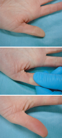
Sepsis is typically diagnosed when there are alterations in conscious level, hypotension, and organ failure, manifesting for example as oliguria, hypoxemia, jaundice, or petechial rash. Delayed diagnosis and intervention are associated with increased morbidity, and a high index of suspicion in all patients with abnormal signs and a possibility of infection should be maintained. Sepsis should be suspected in patients with an unexplained altered mental status, tachypnea with a clear chest and normal oxygenation, or if the clinician's instinct indicates something unusual about a seemingly routine infection.[80]American College of Emergency Physicians (ACEP) Expert Panel on Sepsis. DART: an evidence-driven tool to guide the early recognition and treatment of sepsis and septic shock [internet publication].
https://poctools.acep.org/POCTool/Sepsis(DART)/276ed0a9-f24d-45f1-8d0c-e908a2758e5a
Investigations
Initial investigations cover four purposes:
To identify causative organisms
To evaluate for organ dysfunction
To identify the source of infection
To aid prognosis and the selection of an appropriate level of care.
Priority should be given to those investigations that will help to answer important clinical questions, such as the source of infection and severity of illness. Cultures of blood and other fluids will take 48 to 72 hours to yield sensitivities of causative organisms (if identified), but are far less sensitive if delayed until after antimicrobial administration.
Investigations to identify causative organisms:
Blood cultures should be taken immediately, and preferably before antibiotics are started, provided their sampling will not delay administration of antibiotics.[10]National Institute for Health and Care Excellence. Suspected sepsis: recognition, diagnosis and early management. March 2024 [internet publication].
https://www.nice.org.uk/guidance/ng51
[70]Yealy DM, Mohr NM, Shapiro NI, et al. Early care of adults with suspected sepsis in the emergency department and out-of-hospital environment: a consensus-based task force Report. Ann Emerg Med. 2021 Jul;78(1):1-19.
https://www.annemergmed.com/article/S0196-0644(21)00117-7/fulltext
http://www.ncbi.nlm.nih.gov/pubmed/33840511?tool=bestpractice.com
[82]O'Grady NP, Alexander E, Alhazzani W, et al. Society of Critical Care Medicine and the Infectious Diseases Society of America guidelines for evaluating new fever in adult patients in the ICU. Crit Care Med. 2023 Nov 1;51(11):1570-86.
https://journals.lww.com/ccmjournal/fulltext/2023/11000/society_of_critical_care_medicine_and_the.13.aspx
http://www.ncbi.nlm.nih.gov/pubmed/37902340?tool=bestpractice.com
Ideally, at least one set should be taken percutaneously, and one set from any vascular access device that has been in situ for more than 24 hours.[83]Weinstein MP, Reller RB, Murphy JR, et al. The clinical significance of positive blood cultures: a comprehensive analysis of 500 episodes of bacteremia and fungemia in adults. I. Laboratory and epidemiologic observations. Rev Infect Dis. 1983 Jan-Feb;5(1):35-53.
http://www.ncbi.nlm.nih.gov/pubmed/6828811?tool=bestpractice.com
[84]Diament D, Salomão R, Rigatto O, et al. Guidelines for the treatment of severe sepsis and septic shock: management of the infectious agent - diagnosis. Rev Bras Ter Intensiva. 2011 Jun;23(2):134-44.
https://www.scielo.br/pdf/rbti/v23n2/en_a05v23n2.pdf
http://www.ncbi.nlm.nih.gov/pubmed/25299713?tool=bestpractice.com
Other cultures (e.g., of sputum, cerebrospinal fluid [CSF], pleural fluid, joint fluid, stool, and urine) should be taken as clinically indicated.
If no localizing signs are present, systematic examination and culture of all potential sites of infection, including wounds, catheters, prosthetic implants, epidural sites, and pleural or peritoneal fluid, as indicated by the clinical presentation and history, is required.
If meningitis is suspected (e.g., headache, photophobia, neck stiffness, vomiting), a lumbar puncture (LP) for CSF microscopy and culture should be performed. A computed tomography (CT) scan prior to performing an LP to exclude elevated intracranial pressure is required if there is any clinical suspicion of this.
If an enclosed collection such as an abscess or empyema is suspected, it is recommended that this be drained and cultured early in the course of the illness (within 6-12 hours following identification).[3]Evans L, Rhodes A, Alhazzani W, et al. Surviving sepsis campaign: international guidelines for management of sepsis and septic shock 2021. Crit Care Med. 2021 Nov 1;49(11):e1063-143.
https://journals.lww.com/ccmjournal/fulltext/2021/11000/surviving_sepsis_campaign__international.21.aspx
http://www.ncbi.nlm.nih.gov/pubmed/34605781?tool=bestpractice.com
[85]Jimenez MF, Marshall JC. Source control in the management of sepsis. Intensive Care Med. 2001;27(suppl 1):S49-62.
http://www.ncbi.nlm.nih.gov/pubmed/11307370?tool=bestpractice.com
Intubated patients in whom there is a suspicion of pneumonia should have tracheal aspirates, bronchoalveolar lavage, or protected brush specimens taken.
Evaluation for organ dysfunction:
Baseline assessment should include liver function tests (notably bilirubin), a complete blood count (with differential), coagulation (international normalized ratio, activated partial thromboplastin time), serum creatinine, and blood urea nitrogen.
Serum electrolytes and glucose are frequently abnormal, and should be measured at baseline and regularly until the patient improves.
Elevated serum lactate highlights tissue hypoperfusion, and is assessed using a blood gas sample.[10]National Institute for Health and Care Excellence. Suspected sepsis: recognition, diagnosis and early management. March 2024 [internet publication].
https://www.nice.org.uk/guidance/ng51
In practice, a venous blood gas sample is generally used, as it is generally easier and quicker to obtain compared with an arterial blood gas (ABG).
Markers of inflammation, including C-reactive protein (CRP) and procalcitonin, are of use in determining clinical progress and response to therapy. Serial measures of procalcitonin can be useful to guide the decision on when to discontinue antibiotics, alongside clinical evaluation.[3]Evans L, Rhodes A, Alhazzani W, et al. Surviving sepsis campaign: international guidelines for management of sepsis and septic shock 2021. Crit Care Med. 2021 Nov 1;49(11):e1063-143.
https://journals.lww.com/ccmjournal/fulltext/2021/11000/surviving_sepsis_campaign__international.21.aspx
http://www.ncbi.nlm.nih.gov/pubmed/34605781?tool=bestpractice.com
[86]Schuetz P, Birkhahn R, Sherwin R, et al. Serial procalcitonin predicts mortality in severe sepsis patients: results from the Multicenter Procalcitonin MOnitoring SEpsis (MOSES) Study. Crit Care Med. 2017 May;45(5):781-9.
https://journals.lww.com/ccmjournal/fulltext/2017/05000/Serial_Procalcitonin_Predicts_Mortality_in_Severe.5.aspx
http://www.ncbi.nlm.nih.gov/pubmed/28257335?tool=bestpractice.com
[87]Kip MM, Kusters R, Ijzerman MJ, et al. A PCT algorithm for discontinuation of antibiotic therapy is a cost-effective way to reduce antibiotic exposure in adult intensive care patients with sepsis. J Med Econ. 2015;18(11):944-53.
http://www.ncbi.nlm.nih.gov/pubmed/26105574?tool=bestpractice.com
Do not perform procalcitonin testing without an established, evidence-based protocol.[88]American Society for Clinical Pathology. Thirty five things physicians and patients should question. Choosing Wisely, an initiative of the ABIM Foundation. Jul 2021 [internet publication].
https://web.archive.org/web/20230316185857/https://www.choosingwisely.org/societies/american-society-for-clinical-pathology
Do not order erythrocyte sedimentation rate (ESR) to detect acute inflammation before a diagnosis has been established; CRP is a more sensitive and specific test for the acute phase of inflammation than ESR.[88]American Society for Clinical Pathology. Thirty five things physicians and patients should question. Choosing Wisely, an initiative of the ABIM Foundation. Jul 2021 [internet publication].
https://web.archive.org/web/20230316185857/https://www.choosingwisely.org/societies/american-society-for-clinical-pathology
Investigations to identify the source of the infection:
The source of infection may be immediately evident; for example, with classical signs and symptoms of pneumonia (purulent sputum, dyspnea, tachypnea, cyanosis) or an acute abdomen (abdominal pain, guarding, distension, tenderness, absent bowel sounds). However, in many patients the origin must be actively sought.
Diagnostic studies may identify a source of infection that requires removal of a foreign body or drainage to maximize the likelihood of a satisfactory response to therapy. Chest x-rays and ultrasound scans can be performed at the bedside.[89]American College of Radiology. ACR appropriateness criteria: sepsis. 2023 [internet publication].
https://acsearch.acr.org/docs/3188086/Narrative
Examinations such as CT scanning require transfer of potentially unstable patients and the benefit should be weighed against the risk.
An ECG should be arranged to help exclude other differential diagnoses, including myocardial infarction, pericarditis, and myocarditis. Sepsis also predisposes to myocardial dysfunction (particularly in septic shock) and arrhythmias (e.g., atrial fibrillation).[10]National Institute for Health and Care Excellence. Suspected sepsis: recognition, diagnosis and early management. March 2024 [internet publication].
https://www.nice.org.uk/guidance/ng51
[90]Merx MW, Weber C. Sepsis and the heart. Circulation. 2007 Aug 14;116(7):793-802.
https://www.ahajournals.org/doi/full/10.1161/circulationaha.106.678359
http://www.ncbi.nlm.nih.gov/pubmed/17698745?tool=bestpractice.com
In patients at risk of, or with symptoms compatible with, bacterial endocarditis, a transthoracic or transesophageal echocardiogram is useful. This is also helpful to differentiate between hypovolemic, cardiac, and septic shock, as well as alternative diagnoses, such as valvular abnormalities, pulmonary embolus, myocardial ischemia (with segmental or global dysfunction), hypovolemia, and pulmonary hypertension. If readily available, an echocardiogram may also be appropriate in patients with sepsis of unknown origin.
Certain investigations carry prognostic value and can help determine the need for critical care:
Lactate measurement is a useful assessment of perfusion once a diagnosis of sepsis has been established. Increasing levels of lactate are associated with increasing levels of anaerobic metabolism. Persistently elevated lactate levels may parallel the degree of hypoperfusion or organ failure. High lactate carries adverse prognostic value if elevated to >2 mmol/L (>18 mg/dL), and still worse outcomes are associated with levels >4 mmol/L (>36 mg/dL).[10]National Institute for Health and Care Excellence. Suspected sepsis: recognition, diagnosis and early management. March 2024 [internet publication].
https://www.nice.org.uk/guidance/ng51
[70]Yealy DM, Mohr NM, Shapiro NI, et al. Early care of adults with suspected sepsis in the emergency department and out-of-hospital environment: a consensus-based task force Report. Ann Emerg Med. 2021 Jul;78(1):1-19.
https://www.annemergmed.com/article/S0196-0644(21)00117-7/fulltext
http://www.ncbi.nlm.nih.gov/pubmed/33840511?tool=bestpractice.com
Lactate clearance (the rate at which lactate is cleared over a period of 6 hours) has been demonstrated to be as useful as more invasive tests, such as central venous oxygen saturation, in determining a patient's response to treatment.[91]Nguyen HB, Rivers EP, Knoblich BP, et al. Early lactate clearance is associated with improved outcome in severe sepsis and septic shock. Crit Care Med. 2004 Aug;32(8):1637-42.
http://www.ncbi.nlm.nih.gov/pubmed/15286537?tool=bestpractice.com
[92]Jones AE, Shapiro NI, Trzeciak S, et al. Lactate clearance vs central venous oxygen saturation as goals of early sepsis therapy: a randomized clinical trial. JAMA. 2010 Feb 24;303(8):739-46.
https://jamanetwork.com/journals/jama/fullarticle/185405
http://www.ncbi.nlm.nih.gov/pubmed/20179283?tool=bestpractice.com
Studies with trauma patients have evaluated lactate levels against Acute Physiology and Chronic Health Evaluation (APACHE) scores and lactate clearance rates and found lactate levels to be inferior in informing the prognosis. However, an APACHE score takes 24 hours to calculate.[93]Billeter A, Turina M, Seifert B, et al. Early serum procalcitonin, interleukin-6, and 24-hour lactate clearance: useful indicators of septic infections in severely traumatized patients. World J Surg. 2009 Mar;33(3):558-66.
http://www.ncbi.nlm.nih.gov/pubmed/19148699?tool=bestpractice.com
An alternative measure is serum procalcitonin levels. In patients with acute respiratory infections (including those with sepsis), procalcitonin-guided antibiotic therapy has been shown to reduce the antibiotic course length, reduce antibiotic-related complications, and reduce the 30-day mortality rate.[94]Schuetz P, Wirz Y, Sager R, et al. Effect of procalcitonin-guided antibiotic treatment on mortality in acute respiratory infections: a patient level meta-analysis. Lancet Infect Dis. 2018 Jan;18(1):95-107.
https://www.thelancet.com/journals/laninf/article/PIIS1473-3099(17)30592-3/fulltext
http://www.ncbi.nlm.nih.gov/pubmed/29037960?tool=bestpractice.com
However, evidence for the prognostic value of procalcitonin alone is unclear, and its use in the identification of sepsis is excluded from many guidelines.[3]Evans L, Rhodes A, Alhazzani W, et al. Surviving sepsis campaign: international guidelines for management of sepsis and septic shock 2021. Crit Care Med. 2021 Nov 1;49(11):e1063-143.
https://journals.lww.com/ccmjournal/fulltext/2021/11000/surviving_sepsis_campaign__international.21.aspx
http://www.ncbi.nlm.nih.gov/pubmed/34605781?tool=bestpractice.com
[95]Shehabi Y, Sterba M, Garrett PM, et al. Procalcitonin algorithm in critically ill adults with undifferentiated infection or suspected sepsis: a randomized controlled trial. Am J Respir Crit Care Med. 2014 Nov 15;190(10):1102-10.
https://www.atsjournals.org/doi/full/10.1164/rccm.201408-1483OC#.V0JdkOQpqZM
http://www.ncbi.nlm.nih.gov/pubmed/25295709?tool=bestpractice.com
[96]Andriolo BN, Andriolo RB, Salomão R, et al. Effectiveness and safety of procalcitonin evaluation for reducing mortality in adults with sepsis, severe sepsis or septic shock. Cochrane Database Syst Rev. 2017 Jan 18;(1):CD010959.
https://www.cochranelibrary.com/cdsr/doi/10.1002/14651858.CD010959.pub2/full
http://www.ncbi.nlm.nih.gov/pubmed/28099689?tool=bestpractice.com
[97]Lam SW, Bauer SR, Fowler R, et al. Systematic review and meta-analysis of procalcitonin-guidance versus usual care for antimicrobial management in critically ill patients: focus on subgroups based on antibiotic initiation, cessation, or mixed strategies. Crit Care Med. 2018 May;46(5):684-90.
http://www.ncbi.nlm.nih.gov/pubmed/29293146?tool=bestpractice.com
Surviving Sepsis Campaign guidelines recommend the use of procalcitonin alongside clinical evaluation to guide decisions on the discontinuation of antibiotics in patients with sepsis, but not for the initiation of antibiotics.[3]Evans L, Rhodes A, Alhazzani W, et al. Surviving sepsis campaign: international guidelines for management of sepsis and septic shock 2021. Crit Care Med. 2021 Nov 1;49(11):e1063-143.
https://journals.lww.com/ccmjournal/fulltext/2021/11000/surviving_sepsis_campaign__international.21.aspx
http://www.ncbi.nlm.nih.gov/pubmed/34605781?tool=bestpractice.com
One multicenter randomized trial of 2760 critically ill adults hospitalized with suspected sepsis found that a daily procalcitonin-guided protocol reduced antibiotic duration safely compared with standard care.[98]Dark P, Hossain A, McAuley DF, et al. Biomarker-guided antibiotic duration for hospitalized patients with suspected sepsis: the ADAPT-Sepsis randomized clinical trial. JAMA. 2025 Feb 25;333(8):682-93.
http://www.ncbi.nlm.nih.gov/pubmed/39652885?tool=bestpractice.com
Changes in procalcitonin levels may occur later than those of lactate, although changes in both markers combined are highly predictive of outcome between 24 and 48 hours.[99]Phua J, Koay ES, Lee KH. Lactate, procalcitonin, and amino-terminal pro-B-type natriuretic peptide versus cytokine measurements and clinical severity scores for prognostication in septic shock. Shock. 2008 Mar;29(3):328-33.
http://www.ncbi.nlm.nih.gov/pubmed/18277855?tool=bestpractice.com
Noninvasive impedance echocardiography has been shown to predict poor outcome if a cardiac index of <2 or reduced mitral annular plane systolic excursion is identified.[100]Napoli AM, Machan JT, Corl K, et al. The use of impedance cardiography in predicting mortality in emergency department patients with severe sepsis and septic shock. Acad Emerg Med. 2010 Apr;17(4):452-5.
https://onlinelibrary.wiley.com/doi/full/10.1111/j.1553-2712.2010.00705.x
http://www.ncbi.nlm.nih.gov/pubmed/20370786?tool=bestpractice.com
[101]Havaldar AA. Evaluation of sepsis induced cardiac dysfunction as a predictor of mortality. Cardiovasc Ultrasound. 2018 Nov 30;16(1):31.
https://cardiovascularultrasound.biomedcentral.com/articles/10.1186/s12947-018-0149-4
http://www.ncbi.nlm.nih.gov/pubmed/30501628?tool=bestpractice.com
Patients suffering from septic shock who have not responded to initial fluid resuscitation will require intensive monitoring and treatment in high dependency units.
Emerging tests
Molecular diagnostic assays are being developed that can test for common pathogens. Such assays allow more rapid organism identification and antimicrobial susceptibility testing than standard blood culture systems.[109]US Food and Drug Administration. FDA allows marketing of test to identify organisms that cause bloodstream infections and provide antibiotic sensitivity results. Feb 2017 [internet publication].
https://www.fda.gov/news-events/press-announcements/fda-allows-marketing-test-identify-organisms-cause-bloodstream-infections-and-provide-antibiotic
[110]Pancholi P, Carroll KC, Buchan BW, et al. Multicenter evaluation of the Accelerate PhenoTest BC kit for rapid identification and phenotypic antimicrobial susceptibility testing using morphokinetic cellular analysis. J Clin Microbiol. 2018 Apr;56(4):e01329-17.
https://jcm.asm.org/content/56/4/e01329-17.long
http://www.ncbi.nlm.nih.gov/pubmed/29305546?tool=bestpractice.com
[111]Blauwkamp TA, Thair S, Rosen MJ, et al. Analytical and clinical validation of a microbial cell-free DNA sequencing test for infectious disease. Nat Microbiol. 2019 Apr;4(4):663-74.
http://www.ncbi.nlm.nih.gov/pubmed/30742071?tool=bestpractice.com