Images and videos
Images


Ischemic bowel disease
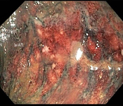
Colonoscopy: mucosal sloughing and likely nonviable colon
From the collection of Dr Jennifer Holder-Murray; used with permission
See this image in context in the following section/s:

Ischemic bowel disease
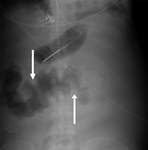
Plain abdominal x-ray: shows marked wall thickening of the transverse colon compatible with the finding of thumbprinting (white arrows)
From the collection of Dr Amir Bastawrous; used with permission
See this image in context in the following section/s:
Ischemic bowel disease
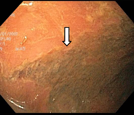
Colonoscopy: demarcation between ischemic and normal colon
From the collection of Dr Jennifer Holder-Murray; used with permission
See this image in context in the following section/s:

Ischemic bowel disease
Comparison of symptoms/signs and investigations for the three types of ischemic bowel disease
Designed by BMJ Knowledge Centre, with input from Dr Amir Bastawrous
See this image in context in the following section/s:
Ischemic bowel disease
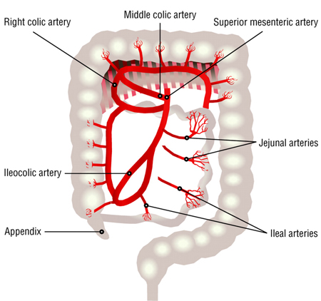
Distribution of blood supply to the small intestine and colon from the superior mesenteric artery, branches of which include the middle, right, and ileocolic arteries as well as jejunal and ileal arteries and arterioles
BMJ 2003; 326 doi: 10.1136/bmj.326.7403.1372
See this image in context in the following section/s:

Ischemic bowel disease
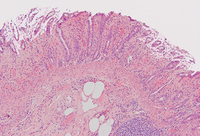
Histopathology of intestinal ischemia
From the collection of Dr Jennifer Holder-Murray; used with permission
See this image in context in the following section/s:

Ischemic bowel disease
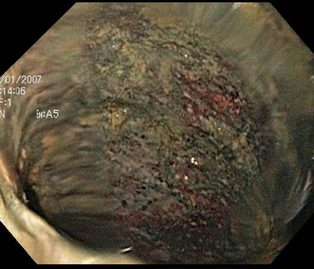
Colonoscopy: denudation of colonic mucosa
From the collection of Dr Jennifer Holder-Murray; used with permission
See this image in context in the following section/s:
Ischemic bowel disease
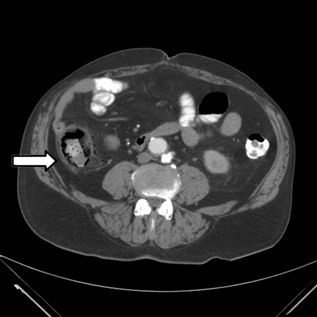
CT scan: colonic thickening with pneumatosis intestinalis
From the collection of Dr Jennifer Holder-Murray; used with permission
See this image in context in the following section/s:
Ischemic bowel disease
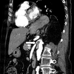
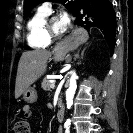
CT angiogram: acute superior mesenteric artery thrombus
From the collection of Dr Jennifer Holder-Murray; used with permission
See this image in context in the following section/s:

Ischemic bowel disease
CT angiography: 3-dimensional reconstruction with superior mesenteric artery stenosis from severe atherosclerotic plaque in a patient on follow-up imaging for endovascular aneurysm repair
From the collection of Dr Jennifer Holder-Murray; used with permission
See this image in context in the following section/s:
Ischemic bowel disease
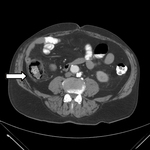
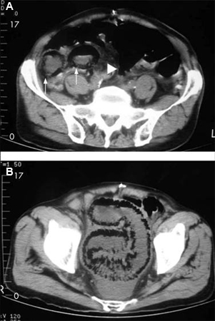
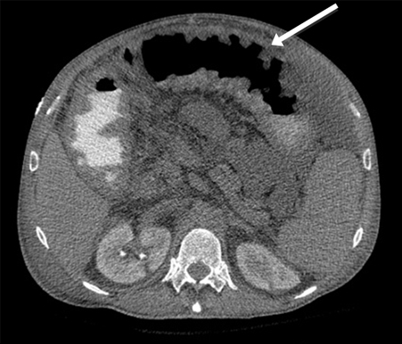
84-year-old man presenting with symptoms suggestive of ischemic bowel disease: (A) Abdominal CT revealing a massive circumferential and band-like air formation as intestinal pneumatosis (arrows) and pronounced edema of mesenteric fat (arrowhead) around necrotic bowel loops; (B) Another slice of abdominal CT showing long segmental pneumatosis of the small bowel
Lin I, Chang W, Shih S, et al. Bedside echogram in ischaemic bowel. BMJ Case Reports. 2009:bcr.2007.053462
See this image in context in the following section/s:

Ischemic bowel disease
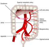
Distribution of blood flow to the colon originating from the inferior mesenteric artery, branches of which include the left colic, marginal, and sigmoid arteries and supply the left colon and superior portion of the rectum
BMJ 2003; 326 doi: 10.1136/bmj.326.7403.1372
See this image in context in the following section/s:
Ischemic bowel disease
CT scan: circumferential wall thickening of the transverse colon; white arrow shows thumbprinting
From the collection of Dr Amir Bastawrous; used with permission
See this image in context in the following section/s:
Videos
 Venepuncture and phlebotomy: animated demonstration
How to take a venous blood sample from the antecubital fossa using a vacuum needle.
 How to perform an ECG: animated demonstration
How to record an ECG. Demonstrates placement of chest and limb electrodes.
 Central venous catheter insertion: animated demonstration
Ultrasound-guided insertion of a non-tunnelled central venous catheter (CVC) into the right internal jugular vein using the Seldinger insertion technique.
 Peripheral intravascular catheter: animated demonstration
How to insert a peripheral intravascular catheter into the dorsum of the hand.
 Female urethral catheterization: animated demonstration
How to insert a urethral catheter in a female patient using sterile technique.
 Male urethral catheterization: animated demonstration
How to insert a urethral catheter in a male patient using sterile technique.
 Nasogastric tube insertion animated demonstration
How to insert a fine bore nasogastric tube for feeding.
Use of this content is subject to our disclaimer