Approach
Smallpox has been eradicated and is only likely to re-emerge via an accident at the known repositories, if a misplaced vial of variola virus from the smallpox era is mishandled, or as the result of bioterrorism.
It is important to keep possible differential diagnoses in mind when assessing a patient presenting with an acute febrile illness and a rash, as they are far more likely to be the cause. A definitive diagnosis of smallpox can only be made if the appropriate clinical case definition has been met, and the diagnosis has been confirmed by laboratory testing.
Smallpox is a notifiable condition and one case is considered an outbreak. Immediately report suspected cases to national or local public health authorities, regardless of whether you are also exploring other potential diagnoses. A highly suspicious or true smallpox event must be reported urgently to the World Health Organization in order to generate an immediate global public health response.
Infection prevention and control
Immediately contact your regional infectious disease unit if there is a clinical suspicion of smallpox.
This will trigger procedures to be activated for the safe transfer of the patient to a negative-pressure isolation facility and the notification of the public health team.
It is important to keep records of everyone who has been in close contact with the symptomatic patient (e.g., household contacts, paramedical and medical staff).
All suspected cases should be managed by experts, including public health officials, to prevent a potential emergency situation.
Follow your local infection prevention and control protocols.
Standard, contact, droplet, and airborne precautions are recommended.
Ensure you have appropriate personal protective equipment (PPE) for the assessment of suspected cases. Healthcare workers must wear full PPE including an N95 face mask to protect against transmission of infection by the respiratory route and physical contact with the patient or patient’s bodily fluids. Rigorous donning and doffing of PPE should be undertaken in the presence of a trained observer.[24]
Treat all contaminated materials (e.g., linens, hospital gowns) and body fluids/solid waste of patients as potentially infectious. Deceased patients are also considered infectious.
Ideally all personnel likely to be in contact with the patient, bodily fluids, or fomites should have been vaccinated with the smallpox vaccine.
History
In the initial stages of a new smallpox epidemic, there may be no clear history of exposure if there is a surreptitious, deliberate release of the virus. There may be a history of exposure if a worker at one of the smallpox repositories is affected, or if a laboratory worker accidentally encounters the virus in another institution while disposing of archival material. A history of suspicious aerosol delivery or terrorist propaganda claiming a biologic attack may also be present.
Immunocompromised patients, pregnant women, and children are at increased risk of severe disease.[1]
Clinical presentation
The incubation period (range 7-17 days) is followed by a sudden onset of influenza-like symptoms that can last for 2-4 days (prodromal or pre-eruptive period). The patient becomes infectious once the fever develops, and they are most infectious during the first week of illness.[1]
Common symptoms include:[1]
Characteristic rash
Fever
Malaise
Prostration
Chills
Headache
Backache
Myalgia
Pharyngitis
Nausea/vomiting.
Less common symptoms include:[1]
Abdominal pain
Diarrhea
Delirium/confusion
Seizures.
Fever (usually high), malaise, and prostration are usually dramatic.
Infection caused by variola major is the more severe and most common form, with a more extensive rash and higher fever. Infection caused by variola minor (alastrim) is a much less severe disease, and was common in the US when the disease was endemic. However, it is not possible to reliably distinguish between infection with the two variants clinically without laboratory assistance.
Complications including pockmarks or pitted lesions (most commonly on the face), panophthalmitis and blindness from viral keratitis or secondary infection of the eye, encephalitis, arthritis, sepsis, and pneumonia may occur.[25] See Complications.
Physical examination
Shortly after the prodromal period, a characteristic rash develops. As the rash develops, the fever begins to subside. However, the fever may return as the rash progresses.[1]
The rash usually presents 1-3 days after the onset of the acute febrile illness, and typically spreads to all parts of the body within 24 hours. It is preceded by enanthema affecting the oropharynx and tongue 24 hours prior, which often passes unnoticed. The rash may be visible during the prodromal period in pale-skinned patients.
Lesions are all of a similar stage of maturation on any part of the body, and tend to be more concentrated on the face and extremities (centrifugal) rather than on the trunk. It can often affect the palms and soles, and may appear on mucous membranes of the nose and mouth. Lesions may be discrete or confluent.
The lesions simultaneously progress through four stages (macular, papular, vesicular, and pustular) before scabbing over and resolving, typically over a period of 2-4 weeks.
As the rash develops, macules progress to papules within 1-2 days. Papules then progress to vesicles within another 1-2 days. The vesicles are well-circumscribed and located deep in the dermis, so individual vesicles do not readily rupture (unlike varicella and herpes simplex vesicles).
As the rash progresses, vesicles progress to pustules in another 1-2 days. Pustules are generally up to 6 mm in smallpox. Pustules subsequently umbilicate before scabbing over and gradually separating after approximately 2 weeks. Once the scab drops off, the person is no longer considered contagious. A depressed scar or areas of pigmentation may remain, most notably on the face.
Generally speaking, lesions are less numerous with variola minor infection and more numerous with variola major infection.
[Figure caption and citation for the preceding image starts]: Classic maculopapular smallpox rash on torso and armCDC/Jean Roy [Citation ends].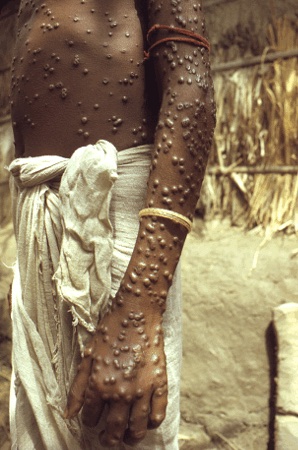
Smallpox may not always present with a classical rash. Typical smallpox occurs in about approximately 85% to 90% of cases, but other presentations are possible.[1][25]
Modified-type smallpox: occurs in approximately 2% of cases, with smaller, more rapidly evolving and more superficial lesions. Usually occurs in those who are previously vaccinated, and is rarely fatal.
Malignant (flat-type) smallpox: occurs in approximately 7% of cases, with relentlessly evolving flat lesions that eventually merge. Raised vesicles or pustules are rarely seen, and the lesions remain flush with the skin with very little vesicular fluid. This type of smallpox usually affects unvaccinated children and carries a serious prognosis (97% mortality in the absence of prior vaccination). It is preceded by a severe prodromal illness with a marked enanthem.
Hemorrhagic smallpox: occurs in approximately 3% of cases, with epistaxis, hematuria, subconjunctival hemorrhages, blood in feces, and metrorrhagia. There may be a hemorrhagic rash and a hemorrhagic enanthem. Fatality usually occurs around day 6. Hemorrhagic smallpox causes significant diagnostic difficulty because of the atypical rash and other features. Occasionally hemorrhagic features occur late in the course of classical smallpox.
Variola sine eruptione: may rarely present with no rash at all, with a mild or even asymptomatic infection. The virus gains little more than a foothold because of preexisting immunity from vaccination or maternal antibodies.
Other factors may affect disease severity (e.g., pregnancy, immunocompromised), resulting in more severe disease in terms of the number and confluency of vesicles and pustules, and a poorer prognosis.
Initial investigations
Definitive diagnosis of smallpox requires laboratory confirmation. Polymerase chain reaction (PCR) is the preferred laboratory test given its accuracy and sensitivity. Testing is available at regional public health laboratories.
The recommended specimen type is skin lesion material, including swabs of lesion exudate, roofs from more than one lesion, or lesion crusts.
Collect other diagnostic material as dictated by the reference laboratory protocol, such as oropharyngeal swabs, ethylenediamine tetra-acetic acid (EDTA) blood, clotted blood for serum separation, or urine.
Collect, label, package, and send specimens according to local or national protocols. Specimens should be collected by vaccinated healthcare workers wearing appropriate PPE. Notify the laboratory of the possibility of smallpox prior to sending specimens. There are local protocols in place for the safe handling of these specimens in the laboratory and onward transport of virologic materials to the reference laboratory. Package samples for testing for other infections separately.
Order routine blood tests, including:
Complete blood count
BUN and electrolytes
Liver function tests
Venous lactate
Clotting screen (if hemorrhagic smallpox is suspected).
Judicious selection of investigations is important in order to reduce risk of transmission to laboratory workers and other healthcare personnel.
Other investigations
Other methods of smallpox diagnosis (e.g., electron microscopy, immunohistochemistry, serologic testing) are either not fully confirmatory or impractical; therefore, PCR is the test of choice.
Order a blood culture in patients with suspected infection (while the patient is in isolation and prior to antibiotic therapy) if there is suspicion of bacterial superinfection of cutaneous lesions or bacterial infection in a very sick patient.
Always rule out coinfection with malaria in any febrile patient who has been to a malaria-endemic area, especially in the 3 weeks prior to the onset of fever. An antigen detection test poses less of an infection hazard to laboratory staff than preparation of thick and thin films.
Differential diagnosis
The most important illness to differentiate from smallpox is acute varicella-zoster (chickenpox). Chickenpox has a longer incubation period (usually 14-21 days), and lesions tend to develop on the trunk and then progress to the face and extremities, rarely affecting the palms and soles. The rash progresses at varying rates.
The Centers for Disease Control and Prevention has produced a diagnostic algorithm that provides a standard method for evaluating patients with acute, severe vesicular or pustular rash illness by giving clinical clues for differentiating smallpox from other diseases. It relies on determining the patient's risk for smallpox based on defined major and minor clinical criteria. The clinical and public health response then depends on the patient’s risk category (high, moderate, low). Testing for smallpox is only recommended in high-risk patients (i.e., those who have a febrile prodrome and classic smallpox lesions and the lesions are at the same stage of development).
Use of this content is subject to our disclaimer