Approach
The diagnosis is suggested by the characteristic history and exam findings of:
Raynaud phenomenon
Sclerodactyly
Skin thickening proximal to metacarpophalangeal joints (in addition to sclerodactyly)
GERD
Digital ulcers
Telangiectasias
Pulmonary involvement.
History
The history should include inquiry about risk factors that are strongly associated with the development of scleroderma, including a family history of the disease or immune dysregulation (e.g., presence of antinuclear antibody [ANA]).
Patients often present with diffuse hand swelling. This is usually associated with hand stiffness that can be misinterpreted as synovitis. The hand swelling is often worse in the morning and improves as the day wears on, only to recur the following morning. Hand swelling can be accompanied by foot swelling, which can prompt investigations into heart failure, renal impairment, or nephrotic syndrome. Raynaud phenomenon is the most common feature of vascular disease and is another initial feature in most people with scleroderma.
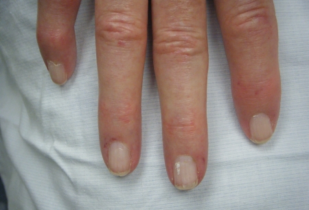
As the initial phase of swelling resolves, the patient may notice skin thickening and the diagnosis becomes more obvious. Patients may complain of loss of function of their hands, particularly the inability to get a tight grasp on objects, as a result of the development of sclerodactyly. This may cause more complaints than skin thickening or tightness. [Figure caption and citation for the preceding image starts]: Hand demonstrating sclerodactyly with finger curling, shiny skin at the fingers, and telangiectasiasFrom the collection of Maureen D. Mayes, University of Texas [Citation ends].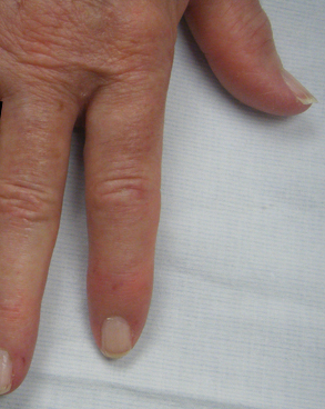
Patients may also initially seek medical attention for heartburn, reflux, dysphagia, or weight loss, and be unaware of the development of sclerodactyly. With further gastrointestinal (GI) involvement there may be symptoms of small bowel disease, including abdominal distension, bloating, and constipation. There may also be melena or symptoms associated with anemia due to chronic GI bleeding (e.g., with gastric antral vascular ectasia [GAVE]). Other early symptoms include fatigue, arthralgias, and myalgias.
An explosive onset with rapid skin progression after onset of Raynaud phenomenon is characteristic of diffuse disease. This may include complaints of shortness of breath, dry cough, or decreased exercise tolerance, indicative of pulmonary and/or cardiovascular involvement. Muscular involvement may or may not occur, with muscular weakness.
Physical exam
Suspicion that Raynaud phenomenon is secondary (e.g., to scleroderma) should prompt referral to a rheumatologist. Features that would suggest secondary Raynaud phenomenon include:[14][15]
Intense episodes, especially those associated with ischemic ulcerations (primary Raynaud disease does not cause ulcers)
Other clinical features suggestive of connective tissue disease (e.g., inflammatory arthritis, abnormal PFTs)
Presence of specific autoantibodies
Evidence of microvascular disease on nail-fold capillaroscopy.
Other typical signs in people presenting with scleroderma include the following:
Tendon friction rubs: palpable, and sometimes audible rub on movement of the ankle, wrist, knee, shoulder, or elbow. They are caused by irritation of the tendon sheath or adjacent tissues and may be seen early in the disease. They are a poor prognostic indicator.
Synovitis: can be difficult to diagnose but joint tenderness on compression or squeezing should be present.
Subcutaneous calcinosis: present as small, localized, hard masses on fingers, forearms, or other pressure points.
Telangiectasias: may be numerous and are commonly located on the face, mucous membranes, and hands. [Figure caption and citation for the preceding image starts]: Telangiectasias on the handFrom the collection of Maureen D. Mayes, University of Texas [Citation ends].
 [Figure caption and citation for the preceding image starts]: Telangiectasias on the fingersFrom the collection of Maureen D. Mayes, University of Texas [Citation ends].
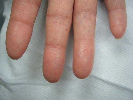
Increased accentuation of the pulmonic component of the second heart sound (S2): suggestive of pulmonary hypertension.
Dry crackles heard on auscultation of primarily the lung bases: suggestive of interstitial lung disease.
An inflammatory arthritis: seen in some patients.
Digital pits, digital ulcerations, and active Raynaud phenomenon. [Figure caption and citation for the preceding image starts]: Digital pits without ulcersFrom the collection of Maureen D. Mayes, University of Texas [Citation ends].
 [Figure caption and citation for the preceding image starts]: Digital tip infarctionFrom the collection of Maureen D. Mayes, University of Texas [Citation ends].
 [Figure caption and citation for the preceding image starts]: Digital tip infarctionFrom the collection of Maureen D. Mayes, University of Texas [Citation ends].
 [Figure caption and citation for the preceding image starts]: Hands demonstrating Raynaud phenomenonFrom the collection of Maureen D. Mayes, University of Texas [Citation ends].
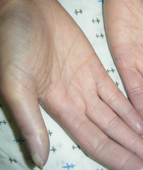
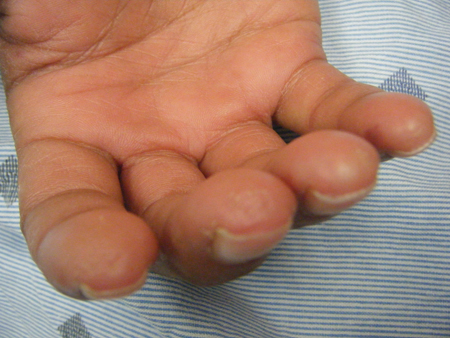
Skin changes: vary in limited and diffuse disease. Limited cutaneous disease is defined by skin thickening distal to the elbows and knees. Involvement of the face is noted in both diffuse and limited disease. Skin changes proximal to the elbows and/or involving the anterior chest and abdomen (in addition to distal skin involvement) are seen in diffuse disease. Sclerodactyly is seen in both types and may be limited to portions of the digits distal to the PIP joint in limited disease. However, it can also result in severe contractures. [Figure caption and citation for the preceding image starts]: Fingers demonstrating sclerodactyly with finger curling, shiny skin at the fingers, and telangiectasiasFrom the collection of Maureen D. Mayes, University of Texas [Citation ends].
 [Figure caption and citation for the preceding image starts]: Hand demonstrating sclerodactyly with finger curling, shiny skin at the fingers, and telangiectasiasFrom the collection of Maureen D. Mayes, University of Texas [Citation ends].[Figure caption and citation for the preceding image starts]: Late-stage sclerodactyly with contractures due to severe skin tighteningFrom the collection of Maureen D. Mayes, University of Texas [Citation ends].
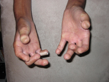 [Figure caption and citation for the preceding image starts]: Late-stage sclerodactyly with contractures due to severe skin tighteningFrom the collection of Maureen D. Mayes, University of Texas [Citation ends].
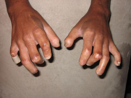
A few patients may present with the abrupt onset of moderate/marked hypertension, which may be malignant hypertension. This is a sign of scleroderma renal crisis, which is seen most commonly in the early stages of diffuse disease.
Investigations
Initial investigations in all patients suspected of having scleroderma include serology test for autoantibodies. Over 90% of patients have a positive ANA. Among this group, there are subsets of mutually exclusive autoantibodies that are associated with distinct clinical phenotypes.
Anti-topoisomerase I (anti-Scl 70) antibody is seen in about 20% of cases and is associated with an increased risk of interstitial lung disease (ILD).
Anti-RNA polymerase III antibody (about 20% of cases) is associated with renal crisis.
Anticentromere antibodies (20% to 25% of cases) are associated with limited skin involvement and a better overall prognosis.
The remaining 40% of patients with clinical scleroderma do not have a known scleroderma-specific autoantibody.
Other initial blood tests include CBC, BUN and serum creatinine, and inflammatory markers such as ESR or C-reactive protein (CRP). Skin biopsy is not generally required in the diagnosis of scleroderma.
Evaluation for pulmonary disease is important as this is the largest cause of mortality in patients with scleroderma. Complete pulmonary function tests (PFTs) with diffusion capacity (including spirometry, lung volumes, and diffusion capacity for carbon monoxide) and a high-resolution chest computed tomography (CT) scan are performed initially.[16] A high-resolution CT scan of the lungs is more sensitive than chest x-ray for early ILD.Complaints of dyspnea, dry cough, or decreased exercise tolerance should prompt further evaluation for pulmonary or cardiac involvement by chest x-ray, ECG, and echo. A yearly PFT (with spirometry, lung volumes, and diffusion capacity) and echo should be done for surveillance in all scleroderma patients. Repeat echocardiogram should be done in those with a high suspicion for pulmonary hypertension or cardiac disease.
Referral for a right-heart catheterization and full evaluation should be done if an echocardiogram finds that the right ventricular systolic pressure (RVSP) is elevated, as echo findings may not be indicative of true pulmonary artery pressures. Estimates of RVSP/pulmonary artery pressure can be obtained from an echocardiogram, based on the tricuspid regurgitation jet (TR jet). If pulmonary hypertension is suspected (declining exercise tolerance in spite of stable PFTs, evidence of right heart failure), referral for right-heart catheterization may be considered even in the face of a normal echo estimate of RVSP.
A barium swallow test can be helpful as an initial investigation to look for features consistent with scleroderma, including dysmotility and reflux. It is also indicated when symptoms of heartburn worsen, or do not improve, with appropriate therapy.
Symptoms and signs of anemia or melena, commonly from chronic GI blood loss from GAVE, require investigation with upper GI endoscopy. CBC may reveal a microcytic anemia.
Patients with scleroderma may develop an inflammatory myositis. In this instance they present with weakness (usually in the proximal muscles). Patients with these symptoms, or those with suspected muscular involvement, require serum muscle enzymes, possibly followed by an EMG/nerve conduction study and muscle biopsy. Scleroderma myopathy may also occur but is detected by elevated muscle enzymes with no muscle weakness.
Use of this content is subject to our disclaimer