Approach
In adult patients, the diagnosis of RTA is more often made after the observation of unexplained, incidental laboratory findings in blood and urine acid-base parameters and electrolyte concentrations. In children the diagnosis is commonly identified following investigation for growth retardation, rickets, or failure to thrive. Rarely, patients may present with severe acidosis and Kussmaul breathing due to respiratory compensation.
Presentation
Classic distal RTA can be asymptomatic or patients may have a history of renal calculi or nephrocalcinosis. Muscle weakness may be present due to associated hypokalemia. Hypokalemic quadriparesis has been described. In a study of patients with defined mutations leading to distal RTA, 55.9% had hypokalemia. Interestingly, patients with ATP6V1B1 or ATP6V0A4 mutations were noted to have more severe hypokalemia than patients with the SLC4A1 mutation.[27]
Hereditary distal RTA usually presents in childhood with failure to thrive and growth retardation. Specific symptoms may include constipation, polydipsia and polyuria, vomiting, dehydration, and anorexia. In some cases rickets, osteomalacia, nephrocalcinosis, and nephrolithiasis may develop.[123] Hereditary distal RTA may also present with sensorineural hearing loss.
Patients with hyperkalemic distal RTA often have a history of diabetes or prostatism.
Children with proximal RTA present with growth retardation and may have renal rickets if Fanconi syndrome is present. The evaluation of Fanconi syndrome otherwise often begins with one of two scenarios. In one, the patient has been found to have hyperchloremic metabolic acidosis with bicarbonate wasting after initial observation of low serum bicarbonate; in the other, the patient has been diagnosed with a condition known to be associated with Fanconi syndrome, either a genetic disorder or an acquired condition such as renal rickets. The hypokalemia associated with proximal RTA and Fanconi syndrome may cause muscle weakness.
Past medical history associated with RTA
Diabetic patients with mild chronic kidney disease and autonomic neuropathy are at risk for hyporeninemic hypoaldosteronism and selective aldosterone deficiency and therefore hyperkalemic distal RTA (type IV).[4][58][59]
Primary adrenal insufficiency, when untreated or undertreated, also predisposes to hyperkalemic distal RTA.
Interstitial nephritis associated with systemic lupus erythematosus, chronic active hepatitis, thyroiditis, and renal transplant may lead to RTA.
Primary Sjogren syndrome appears to carry a significant risk for development of distal RTA, and may present with hypokalemic paralysis.[12]
Hereditary fructose intolerance is associated with Fanconi syndrome.
Up to 50% of patients with primary biliary cirrhosis have been reported to have classic distal RTA.
Hereditary distal RTA is more common in Thai and southeast Asian populations.
Ocular abnormalities (cataracts, glaucoma, band keratopathy), growth retardation, impaired intellect, and calcification of basal ganglia have been linked to a rare form of autosomal recessive proximal RTA.
Hyperparathyroidism may result in proximal RTA and partial Fanconi syndrome.
People with inherited metabolic disorders that have been associated with Fanconi syndrome (e.g., Lowe syndrome, Wilson disease, Dent disease, cystinosis, galactosemia, hereditary fructose intolerance, Von Gierke disease) should be evaluated for proximal RTA, glycosuria, phosphaturia, and aminoaciduria.
Drug, toxin, and environmental exposure
A number of drugs are associated with RTA:
Medications that interfere with distal nephron sodium transport including spironolactone, triamterene, trimethoprim, pentamidine, angiotensin-converting enzyme (ACE) inhibitors, and angiotensin II receptor inhibitor therapy
Carbonic anhydrase inhibitors
Cyclosporine
Amphotericin-B
Heparin
Ibuprofen
Ifosfamide
Lamivudine
Antiviral therapy: cidofovir, tenofovir, adefovir.
Toxins associated with RTA:
Heavy metals
Cis-platinum.
Environmental exposure:
Balkan endemic nephropathy.
Investigative approach
The investigation for RTA typically follows two routes.
Discovery of the presence of an abnormally low serum bicarbonate concentration and hyperchloremia.
Recognition of significant risk factors or consequences of RTA, which suggest that RTA may be present: for example, nephrocalcinosis, diabetes, prostatism, growth retardation, and renal calculi.
The diagnosis of all forms of distal RTA ultimately rests on finding an elevated urine pH or decreased urinary ammonium excretion in the presence of hyperchloremic metabolic acidosis.[2][4] In the case of proximal RTA, the diagnosis requires the demonstration of bicarbonate wasting in a patient presenting with hyperchloremic, hypokalemic metabolic acidosis.[2][4] The confirmatory testing for Fanconi syndrome addresses two evaluations:
Proving the diagnosis of proximal RTA
Evaluating the renal excretion of glucose, phosphate, and amino acids.
Laboratory evaluation
The diagnosis of RTA depends entirely on the laboratory evaluation of blood and urine acid-base parameters and electrolyte concentrations.[4]
The diagnosis always requires the simultaneous determination of the arterial pH, PCO₂, and bicarbonate.
Serum bicarbonate, chloride, sodium, and potassium together with the serum anion gap are routinely performed in all patients.
In hyperkalemic distal RTA the measurement of serum aldosterone is also used to differentiate aldosterone deficiency from aldosterone resistance.
The urine pH must be measured by pH electrode or blood gas analyzer; this is performed in all patients with suspected RTA.
Measurement of serum bicarbonate and urine pH is critical in the presence of recurrent nephrolithiasis.
If the urine pH exceeds 5.5 in the presence of hyperchloremic metabolic acidosis, the diagnosis of classic distal RTA may be made.
If urine pH is normally low (<5.5), the urinary ammonium concentration should be estimated by determination of the urine anion gap.
In urine with a pH under 6.0, if the urine chloride concentration exceeds the sum of sodium and potassium concentrations, ammonium is present in the urine.[124][125][126]
If the sum of the urine sodium and potassium concentrations exceeds the urine chloride concentration in an acid urine (pH <6.0), ammonium must be absent from the urine; this indicates hyperkalemic RTA is present.
In cases where the diagnosis is uncertain, or in which distal RTA is incomplete, physiologic tests are used to confirm the diagnosis of distal RTA. Physiologic tests of acidification include:[126][127][128][129]
The response of the urine pH and potassium concentration to furosemide administration, or alternatively the response of urine pH to furosemide and fludrocortisone to confirm diagnosis of hyperkalemic distal RTA.
The measurement of urine pH after ammonium chloride loading to induce acidosis to help confirm incomplete distal RTA as distal RTA; however, this test is unpleasant for patients.
Confirmation of suspected proximal RTA requires bicarbonate infusion and the measurement of the fractional excretion of bicarbonate.
Interpreting arterial pH, bicarbonate, and anion gap
A low serum bicarbonate concentration is seen in both metabolic acidosis and in respiratory alkalosis, so one starts by establishing that the arterial pH is below normal (under 7.37).
If the arterial pH is normal or elevated, the cause of the low serum bicarbonate is the normal adaptation to respiratory alkalosis.
If the arterial pH is low, the next question is whether the serum anion gap is normal or increased.
In all types of RTA, the anion gap is normal, whereas an increased serum anion gap indicates the presence of another type of acidosis.[125][130]
The combination of depressed serum bicarbonate, hyperchloremia, acidemia, and a normal serum anion gap indicates that patient has hyperchloremic non-gap metabolic acidosis, but does not yet prove that the patient has RTA.[124]

How to obtain an arterial blood sample from the radial artery.
Interpreting urine pH and electrolytes
To complete the diagnosis of RTA, one must also show that urinary acidification is impaired (distal RTA), or that bicarbonate reabsorption is grossly inadequate (proximal RTA).
Either of two urinary findings can make the diagnosis of distal RTA:[50][126]
An abnormally high urine pH (>5.5) during acidemia (in classic distal RTA)
Minimal or absent urine ammonium (in hyperkalemic distal RTA).
Urinary ammonium is estimated by measuring the urine anion gap (UAG) or urine osmolal gap.[131] Both of these calculations give qualitative and not quantitative estimation of urinary ammonium.
The UAG is calculated using the equation: urinary cations [sodium + potassium] – urinary anions [chloride].
In the setting of normal kidney function and hyperchloremic metabolic acidosis, the UAG is less than zero.
When the UAG is positive, it indicates low urinary ammonium.
The UAG cannot be used if the urine pH is greater than 7 as this would suggest the presence of bicarbonate and other organic anions in the urine. This kind of scenario can be seen in patients with diabetic ketoacidosis.
Urinary osmolal gap is another method to estimate urinary ammonium. It is calculated using the following equation: measured urinary osmolality – calculated urinary osmolality, and can be written as 2(urinary sodium + urinary potassium) + (urinary urea nitrogen/2.8) + (urinary glucose/18).[132]
The normal range of urine osmolal gap is between 80 to 150 mEq/L.
A higher urine osmolal gap suggests high urinary ammonium. The urinary osmolal gap can be used in the presence of other anions.
Interpreting serum electrolytes
In RTA the serum bicarbonate is low and hyperchloremia is present but this is not diagnostic in the absence of arterial blood gas measurement.
The diagnosis of proximal RTA requires that one prove that the fractional excretion of bicarbonate is 15% or greater when bicarbonate is infused to raise the serum bicarbonate to approximately 20 mEq/L.[4]
Proximal RTA is characterized by hypokalemia, as is classic distal RTA.[35][4]
Hyperkalemia in distal RTA indicates that aldosterone deficiency or resistance is the cause of the problem.[4]
The evaluation of patients who have underlying medical conditions that place them at risk for distal RTA but who do not have overt acidosis or hypobicarbonatemia requires provocative physiologic tests.
[Figure caption and citation for the preceding image starts]: Overview of blood and urine diagnostic tests in renal tubular acidosisCreated by BMJ Knowledge Centre [Citation ends].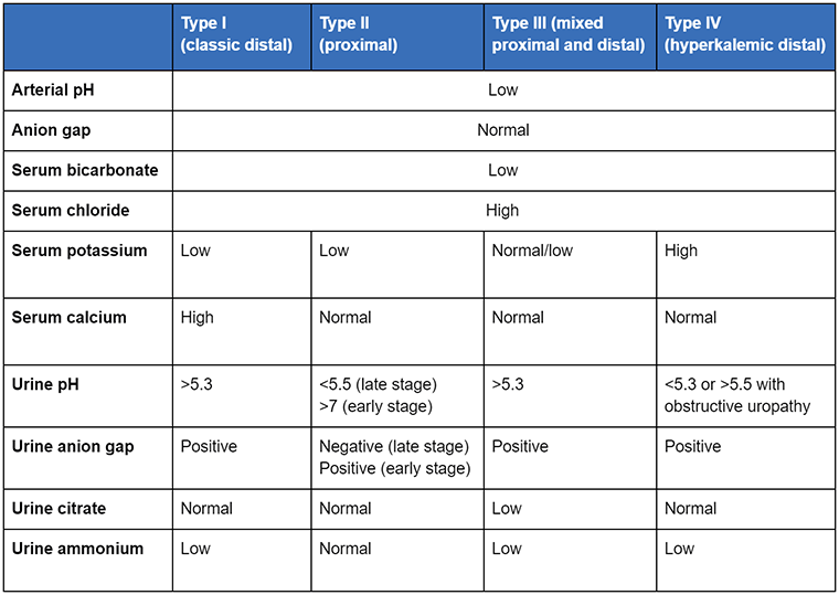
Radiology
Radiologic findings associated with forms of RTA include nephrocalcinosis (in distal RTA), osteopenia, and osteopetrosis, nephrocalcinosis, and cerebral calcifications in hereditary carbonic anhydrase II deficiency.[4][60][61][62][33][34]
Patients with classic distal RTA should have abdominal x-ray or abdominal computed tomography (CT) scan performed to evaluate the kidneys for nephrocalcinosis or the presence of stones. CT scans are superior, but a less costly kidneys, ureter, bladder x-ray may suffice.
Patients with unexplained hyperkalemic distal RTA should be evaluated for urinary tract obstruction by ultrasound, nuclear renal scan, or spiral CT scan. Of these options, ultrasound has the least potential for harm, but is less sensitive than nuclear scans and more operator-dependent than CT. Nuclear scan requires the injection of radioactive tracer, but is the most sensitive test. Spiral CT is neither invasive nor operator-dependent, but is less sensitive than nuclear scan, and is expensive.
There are no radiologic issues of note with proximal RTA or proton backleak. Proximal RTA with Fanconi syndrome may show evidence of renal rickets, but otherwise radiologic findings are rare.
Patients with mutations of the ATP6V0A4 gene may develop enlargement of the vestibular aqueduct, which can be diagnosed by CT or magnetic resonance imaging scan.[14][27][133]
Summary of classic presentations of RTA
Adult patients are often asymptomatic. Very rarely, they may present with Kussmaul breathing (due to severe acidosis and respiratory compensation), and muscular weakness. Paralysis may be present due to hypokalemia.
Laboratory findings together with a past medical history of conditions associated with RTA should prompt further investigation for RTA if necessary.
The combination of growth retardation or renal rickets with hypokalemic, nonanion gap metabolic acidosis and very low serum bicarbonate in a child indicates proximal RTA (type II) or a hereditary distal RTA (type I).[4]
Hypophosphatemia, glycosuria, and excretion of amino acids in the urine is suggestive of Fanconi syndrome with proximal RTA.[4] Rarely, multiple myeloma may be associated with Fanconi syndrome and proximal RTA.[98][99]
Hypokalemic, metabolic acidosis combined with osteopetrosis suggests a mutation of carbonic anhydrase II.[4][33][34] Carbonic anhydrase II deficiency presents with bicarbonate wasting with inability to lower final urine pH.
Hypokalemic nonanion gap metabolic acidosis with sensorineural hearing loss suggests classic distal RTA (type I), but does not distinguish between the major responsible mutations.[15][27][36][37]
In adults, classic distal RTA should be considered in patients with nephrolithiasis and nephrocalcinosis and evidence of hyperchloremic metabolic acidosis.[4][60][61][62][63][64][65] Patients with nephrocalcinosis and nephrolithiasis in the absence of a known cause should be evaluated for incomplete distal RTA.
The acid-base status and serum potassium in adult men with partial urinary tract obstruction should be carefully monitored. Depressed serum bicarbonate here may indicate mild hyperkalemic distal RTA (type IV).[4][54][55]
Use of this content is subject to our disclaimer