Approach
Pattern hair loss is characterized by progressive, patterned hair thinning on the scalp. There is gradual conversion of terminal hair (long, thick, and pigmented) into vellus hair (short, fine, and unpigmented). The condition affects both men and women.
General history and examination
Patients typically present with gradual thinning of hair. History should include onset, duration, and location of hair loss, family history of alopecia, nutrition history, and drug history. There should be no clinical evidence of scarring or inflammation and no signs of tenderness or pruritus. Follicular ostia should be preserved. Past medical history of an autoimmune or inflammatory disorder should be ruled out.
A "hair-pull" test should be conducted. This rapid test is used to measure the telogen hair density. Approximately 50 to 100 hairs are grasped between the thumb and the middle and index fingers and then firmly pulled. The test should be performed on the frontal and occipital areas for comparison. More than 3 hairs pulled is a positive result. In pattern hair loss, the hair-pull test is usually negative (≤3 hairs being pulled).[Figure caption and citation for the preceding image starts]: Hair-pull testFrom the collection of Paradi Mirmirani MD; used with permission [Citation ends].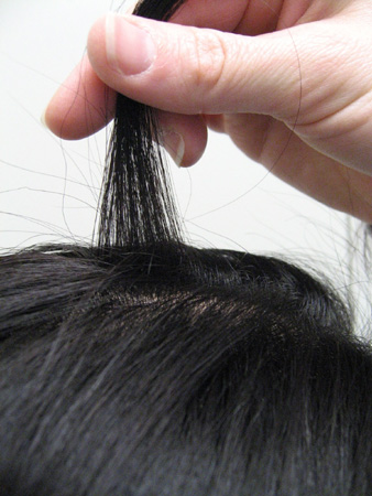
Early onset of hair loss in the vertex area appears to be a marker for early onset of coronary heart disease in people with hypertension and/or dyslipidemia.[6][26] Therefore, additional cardiovascular-related questioning and examination may be appropriate in some patients.
Clinical features in men
A symmetric, progressive, patterned hair loss characterized by bitemporal recession and thinning at the crown (vertex) is seen. The Norwood-Hamilton classification is commonly used to document severity of baldness (types I-VII, based on pattern and severity).[1] The condition can occur at any age, most often following puberty, when androgen hormone levels rise. The first indication is typically a recession of the hairline in the bitemporal areas.[Figure caption and citation for the preceding image starts]: The pattern of androgenetic alopecia in men (after Hamilton and Norwood)From the collection of Robert Haber, MD [Citation ends].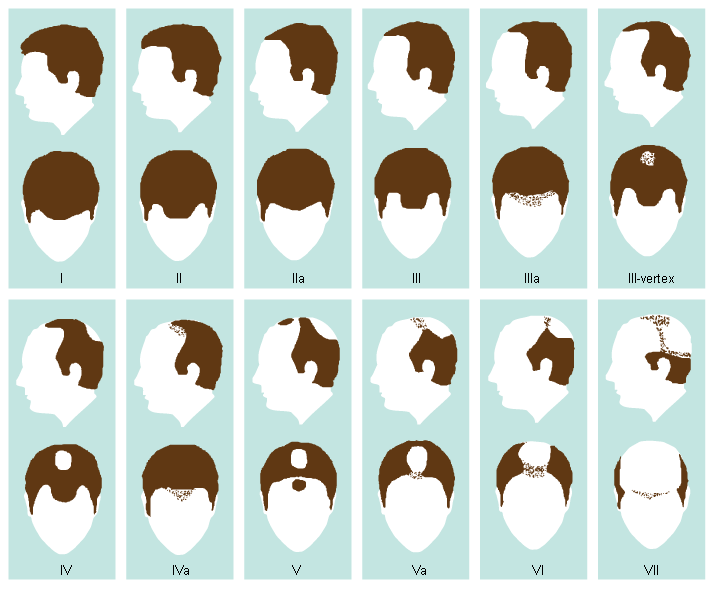
Clinical features in women
The hair loss is more diffuse and less patterned, and female patients typically present with gradual thinning of the central scalp hair, usually over several years.[7][Figure caption and citation for the preceding image starts]: Female-pattern androgenetic alopeciaFrom the collection of Robert Haber, MD [Citation ends].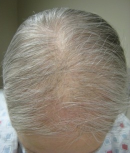 Physical exam shows a widening of the central parting, with diffuse reduction of hair follicles on the frontal and central scalp. The frontal and temporal hairlines are generally retained, but a some women do develop recession of bitemporal hairlines as seen in male-pattern baldness.[7] The Ludwig classification is commonly used to classify severity (types I-III, based on severity).[2] The central part width can also be compared with the occipital part width. Early onset in women suggests pathologic hyperandrogenism.[9][Figure caption and citation for the preceding image starts]: The pattern of pattern hair loss in women (after Ludwig classification)From the collection of Robert Haber, MD [Citation ends].
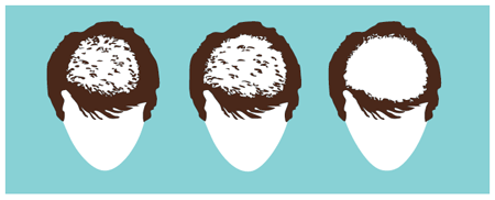
Investigations
It is important to exclude other causes of hair loss.
Laboratory investigations
A history suspicious for autoimmune disease, infection, or anemia in men and women should direct any laboratory testing. This would include CBC with differential, comprehensive metabolic panel, antinuclear antibodies, rapid plasma reagin, and serum ferritin estimation.
In otherwise healthy women, TSH and serum ferritin should be checked.
Women with signs of hyperandrogenism, such as hirsutism on other parts of the body, irregular periods, or severe acne vulgaris, require laboratory analysis of serum dehydroepiandrosterone (DHEA)-sulfate, total and free testosterone levels, and sex hormone-binding globulin.[7] The most common disorder of androgen excess is polycystic ovary syndrome (PCOS). Other causes are adrenal tumors, adrenal hyperplasia, or hyperprolactinemia.[9]
Dermatoscopy (epiluminescence microscopy)/trichoscopy
Usually not required but can be used to examine stage of development of scalp and hairs and to distinguish from other nonscarring forms of alopecia.
Skin biopsy
Usually not required but may be helpful to distinguish from other nonscarring forms of alopecia.[15] Horizontal sections from a scalp biopsy provide important information on the ratio of terminal to vellus hairs and the ratio of hairs in telogen phase (i.e., resting) to anagen phase (i.e., actively growing). There are no significant findings of inflammation. Fibrous streamers, however, can be found around miniaturized hair follicles.
Use of this content is subject to our disclaimer