Approach
The diagnosis is usually a clinical one and may be delayed in regions where tetanus is rarely seen: for example, in the developed world.[40]
History
Immunization status should be clarified. A tetanus-prone wound may be evident; otherwise, patients should be asked about recent injuries, medical interventions including intramuscular injections and obstetric procedures, ear infections, and needle exposure through illicit drug use, acupuncture, or ear piercing.
Wounds or burns that are considered to be tetanus prone include the following:[3][4]
Requiring surgical management but delay in intervention is more than 6 hours
Puncture-type injury or a significant degree of devitalized tissue (especially in contact with soil or manure)
Certain animal bites and scratches
Foreign body-containing wounds
Open fractures
Concomitant systemic sepsis.
Examination
The diagnosis should be strongly suspected if there is trismus (which results in a grimace described as "risus sardonicus," or sardonic smile) and 1 or more of the following: muscle rigidity, spasms, respiratory distress, dysphagia, or autonomic dysfunction (hyperpyrexia, labile blood pressure, cardiac arrhythmias). Intermittent tonic contraction of their skeletal muscles causes intensely painful spasms, which may last for minutes, during which consciousness is retained. The spasms are often triggered by external (noise, light, drafts, physical contact) or internal stimuli and may cause fractures or rhabdomyolysis. Tetanic spasms can produce opisthotonus, board-like abdominal wall rigidity, dysphagia, and apneic periods due to contraction of the thoracic muscles and/or glottal or pharyngeal muscles. During a generalized spasm patients classically arch their back, extend their legs, flex their arms in abduction, and clench their fists. Apnea may be a feature of such a spasm. Autonomic overactivity initially manifests as irritability, restlessness, sweating, and tachycardia. Several days later, hyperpyrexia, cardiac arrhythmias, labile hypertension, or hypotension can occur.
Wounds should be inspected; a patient with no wounds should be examined for other evidence of a portal of entry for spores.[Figure caption and citation for the preceding image starts]: TrismusFrom the collections of Nicholas J. Beeching and Christopher M. Parry [Citation ends].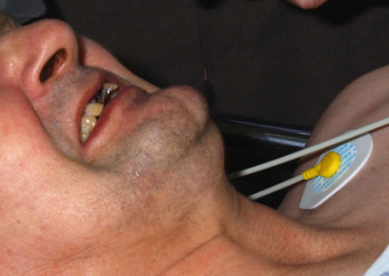 [Figure caption and citation for the preceding image starts]: TrismusFrom the collections of Nicholas J. Beeching and Christopher M. Parry [Citation ends].
 [Figure caption and citation for the preceding image starts]: OpisthotonusFrom the collections of Nicholas J. Beeching and Christopher M. Parry [Citation ends].
 [Figure caption and citation for the preceding image starts]: Observation chart illustrating autonomic dysfunction with extreme fluctuation in blood pressureFrom the collections of Nicholas J. Beeching and Christopher M. Parry [Citation ends].
Laboratory tests
The diagnosis is clinical, but laboratory tests are available to support or confirm the diagnosis. Treatment should never be delayed to wait for laboratory results.
Tetanus toxin can be detected in serum, confirming a clinical diagnosis. Samples should be collected before tetanus immune globulin (TIG) treatment. Absence of toxin in the serum does not exclude the clinical diagnosis.[39]
Clostridium tetani may be detected in the infection site by direct polymerase chain reaction and using strict anaerobic culture methods on wound tissue or swabs. A negative result does not exclude tetanus.[39] A positive wound culture does not indicate whether the organism produces a toxin. The bacteria may be present without causing tetanus in patients with protective immunity.[19]
Demonstration of low levels of tetanus toxin antibodies in serum can support but not confirm a clinical diagnosis. Samples should be taken before TIG treatment, but treatment of clinical tetanus should not be delayed waiting for laboratory result.[39] Conversely, severe and fatal tetanus has been reported in patients with protective antibody levels.[41]
Drug samples or paraphernalia can be tested for the presence of spores after discussion with the relevant health authority.
If clinical diagnosis is uncertain additional investigations may be performed to exclude alternative diagnoses (e.g., hypocalcemia, meningitis). In tetanus infection cerebrospinal fluid findings are usually normal, although cerebrospinal fluid protein - immunoglobulin G - may be slightly elevated.[25][42] Electroencephalogram is normal while electromyogram may be normal or show nonspecific changes.[1]
Use of this content is subject to our disclaimer