Approach
The diagnosis of styes (hordeola) and chalazia requires only history and physical exam. No diagnostic tests are required or useful in their diagnosis. In the acute setting, it may be difficult to differentiate an internal hordeolum from a chalazion. However, because they initially require common treatment modalities that are relatively benign, this differentiation is of limited clinical utility.
External hordeolum
Patients typically present with an external hordeolum complaining of acute onset of unilateral eyelid pain. Pain and swelling involve either the upper or lower lid and are generally well localized to a discrete area, which is tender to palpation. Tenderness is localized at the lid margin in the case of an external hordeolum, giving the classic appearance of the stye. The ciliary glands of Zeis and Moll are typically involved.[13] The hordeolum appears as a pustule that points toward the lid margin. The lid itself may be slightly swollen with some erythema around the involved area. A diffusely swollen lid with marked erythema should prompt the clinician to consider other diagnoses.[1][2][13][Figure caption and citation for the preceding image starts]: Left lower lid external hordeolumCreative Commons CC0 1.0 Universal Public Domain (https://creativecommons.org/publicdomain/zero/1.0/) [Citation ends].
Internal hordeolum
Patients with an internal hordeolum also present with acute pain and swelling of the eyelid. Infection of the meibomian gland, which sits deeper in the lid within the tarsal plate, is responsible for internal hordeola.[2][13] Infection may cause obstruction of the meibomian gland, which is a relatively long gland and has an orifice at the lid margin. If it is obstructed, there may be no notable pathology at the lid margin. Tenderness and erythema may be more diffusely located in the lid. The hordeolum may point toward the conjunctival surface and be diagnosed by everting the lid to reveal a small pustule, which is the hallmark of an internal hordeolum.[2][13] If the gland is infected but not obstructed, physical exam may be similar, without the internal pustule, but with pain and tenderness at the lid margin, as with external hordeola. As the treatment is the same for both internal and external hordeola, this differentiation is not of great clinical concern.[1][13]
[Figure caption and citation for the preceding image starts]: Left upper lid internal hordeolum with limited cellulitisGupta A, Stacey S, Amissah-Arthur KN. Eyelid lumps and lesions. BMJ. 2014;348:g3029. [Citation ends].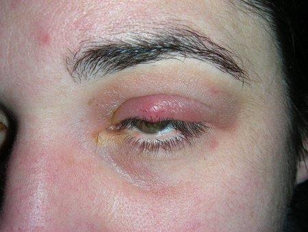
Chalazion
In contrast to hordeola, chalazia typically present in a more indolent fashion. Chalazia result from a localized inflammatory reaction to sebum rather than an acute infectious process. When they arise, they may be confused with an internal hordeolum, lacking any pustule at the lid margin. However, chalazia are nontender nodules and the surrounding skin typically lacks any erythema or signs of an acute bacterial infection. If chalazia persist, chronic skin changes may appear overlying the nodule.[1][6][13]
[Figure caption and citation for the preceding image starts]: Lower lid chalazion of the left eyeGupta A, Stacey S, Amissah-Arthur KN. Eyelid lumps and lesions. BMJ. 2014;348:g3029. [Citation ends].
Pitfalls in diagnosis
While the diagnosis of chalazia and hordeola is typically straightforward, there are several entities with which they may be confused. A complete history should be obtained to identify any contributory factors or point to a differential diagnosis (e.g., visual changes, eye trauma, immunocompromised status, infections, cancer history, travel history). Specifically, pain and swelling of the lid without a discrete visible pustule at the lid margin or conjunctival surface should prompt the clinician to consider other diagnoses. Periorbital (preseptal) and orbital (septal) cellulitis are more diffuse processes and should be considered if the exam lacks a pustule or papule, as this has significant treatment implications. Such patients usually have more diffuse swelling, edema, and erythema of the lid and periorbital region.[4][13]
Ophthalmology referral for possible malignancy is recommended for ulceration and destructive changes of the eyelid margin and in patients with chronic or recurrent chalazia.[7][8][14] Consider early referral for young children with large chalazion due to risk of amblyopia.[7]
Imaging
Computed tomography (CT) scanning of the face and orbits is not useful in diagnosing hordeola or chalazia. However, if alternative diagnoses cannot be differentiated clinically, CT scanning may be appropriate to confirm the diagnosis and exclude other, more serious pathology such as orbital cellulitis.[13]
Use of this content is subject to our disclaimer