Images and videos
Images


Raynaud phenomenon
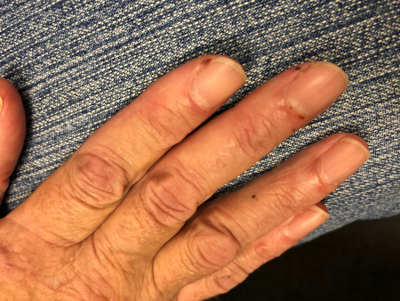
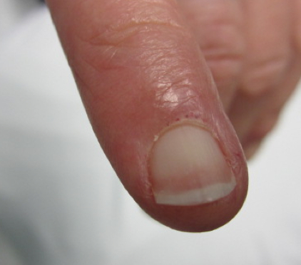
Digital pits on the 3rd finger of the left hand
From the personal collection of Dr Janet Pope; used with permission
See this image in context in the following section/s:
Raynaud phenomenon
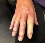
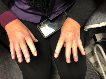
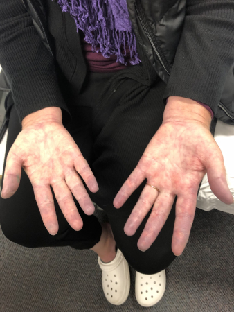
Swollen/puffy fingers in a patient with secondary Raynaud phenomenon (RP)
From the personal collection of Dr Janet Pope; used with permission
See this image in context in the following section/s:
Raynaud phenomenon
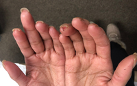
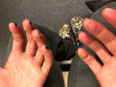
Digital infarcts and nailfold changes
From the personal collection of Dr Janet Pope; used with permission
See this image in context in the following section/s:
Raynaud phenomenon
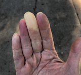
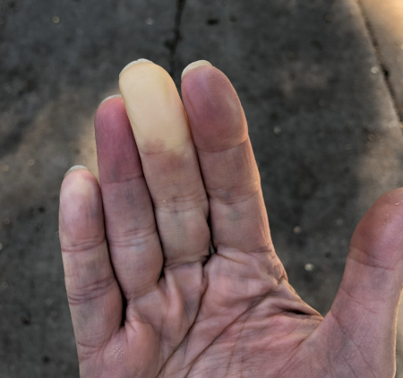
Well-demarcated pallor of the 3rd finger in a patient with primary Raynaud phenomenon (RP)
From the personal collection of Dr Janet Pope; used with permission
See this image in context in the following section/s:
Raynaud phenomenon
Visible dilated capillaries at the nailbed
From the personal collection of Dr Janet Pope; used with permission
See this image in context in the following section/s:

Raynaud phenomenon
Raynaud phenomenon (RP) and inflammatory arthritis with sclerodactyly distal to the proximal interphalangeal joints
From the personal collection of Dr Janet Pope; used with permission
See this image in context in the following section/s:
Raynaud phenomenon
Cyanotic Raynaud phenomenon (RP) in a patient with mixed connective tissue disease
From the personal collection of Dr Janet Pope; used with permission
See this image in context in the following section/s:

Raynaud phenomenon
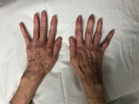
Secondary Raynaud phenomenon (RP) with sclerodactyly
From the personal collection of Dr Janet Pope; used with permission
See this image in context in the following section/s:

Raynaud phenomenon
Digital ulcer in a patient with secondary Raynaud phenomenon (RP)
From the personal collection of Dr Janet Pope; used with permission
See this image in context in the following section/s:
Use of this content is subject to our disclaimer