Urgent considerations
See Differentials for more details
Some of the conditions causing hypotension may present with shock, requiring urgent lifesaving treatment. Shock is the life-threatening failure of adequate oxygen delivery to the tissues, due to decreased blood supply or increased demand, resulting in decreased end-organ oxygenation. If left untreated, shock results in end-organ damage and death. Tissue hypoperfusion may occur without hypotension but, in clinical practice, shock is commonly diagnosed when both arterial hypotension and organ dysfunction are present.[18] Tachycardia, altered cognition or a reduced level of consciousness, diaphoresis, and oliguria are all pointers to the presence of shock.
Airway, breathing, and circulation (ABC) measures are first priority in all critically ill patients. Stabilization of airway and breathing is a priority, followed by restoration of circulating volume and perfusion of peripheral tissues.
Administration of supplemental oxygen with continuous pulse oximetry may be adequate to maintain oxygen saturation. In more severe cases, intubation and ventilation may be required. Start supplemental oxygen therapy through a reservoir mask with a flow rate of 15 L/min for critically ill patients, until reliable pulse oximetry readings are available. Further oxygen therapy should be guided by pulse oximetry, keeping oxygen saturation between 94% and 98%. Patients at risk of hypercapnia may need controlled oxygen therapy to maintain oxygen saturation between 88% and 92%.[19]
Early and frequent monitoring of blood pressure is an important part of perfusion assessment, and an initial fluid bolus should be administered to patients with evidence of intravascular hypovolemia.
All patients require an ECG and, in more severe cases, continuous cardiac monitoring may be indicated. Specific urgent measures are required in specific conditions, listed below.
Hypovolemic shock
Severe dehydration, due to nonhemorrhagic losses (e.g., severe diarrhea and vomiting):
Urgent ABC assessment and support is required. In most cases, intravenous isotonic saline is the initial fluid used.
Monitoring by clinical exam of volume status, BP and central venous pressure (CVP) measurement, combined with close monitoring of electrolytes and renal parameters are important.[9] Aggressive fluid administration is guided by achievement of normal CVP (8-12 cm H2O).
Particular care concerning the rate of fluid replacement is required in older adults and in patients with a history of cardiac failure.
Hemorrhage (e.g., due to gastrointestinal bleed, retroperitoneal bleed, ruptured abdominal aortic aneurysm, ruptured ectopic pregnancy, trauma):
Urgent ABC assessment and support is required. If bleeding is severe, it is typically appropriate to mobilize a multidisciplinary team, including emergency medicine physicians, surgeons, and radiologists.
Intravenous saline is administered initially, and often continued following commencement of blood transfusion.
Blood transfusion is an immediate priority. In the setting of acute large hemorrhage, transfusion of 2 to 4 units is often necessary. The presence of ongoing tachycardia, hypotension, and other signs of shock, or lack of serum hemoglobin response to blood transfusion, are all markers of ongoing internal or occult hemorrhage. Clinical response, pulse, BP, CVP, renal output, complete blood count, coagulation, and electrolytes should be closely monitored. Results from one multicenter cohort study suggested that a systolic BP <110 mmHg is a marker of poor outcomes and higher mortality in blunt trauma patients.[20] Trauma specialists suggest aiming for controlled hypotension (systolic <90 mmHg) in penetrating trauma.
Fresh frozen plasma or coagulation factor concentrates are often necessary when coagulopathy is known or suspected to be present (refer to local hospital guidelines).[21][22][23] Patients on therapeutic anticoagulation should have reversal of anticoagulation, in line with local hospital guidelines. This may include administration of intravenous vitamin K and prothrombin complex concentrate for reversal of anticoagulation with warfarin, protamine for reversal of anticoagulation with heparin, or specific reversal agents for direct oral anticoagulants where available.[24] This may be required following massive transfusion or in patients with upper gastrointestinal bleeding secondary to chronic liver disease.
Antifibrinolytics (such as tranexamic acid) should be considered in all patients as soon as possible with acute severe hemorrhage as they have been shown to increase survival. Among patients with postpartum or traumatic bleeding, immediate treatment with tranexamic acid was found to greatly increase the odds of survival, with the survival benefit decreasing by about 10% for every 15 minutes of treatment delay until 3 hours, after which there was no benefit.[25]
Definitive action to correct the source of hemorrhage is usually only possible when the patient's condition has been stabilized.
Upper or lower gastrointestinal endoscopies should follow initial efforts to stabilize the patient, if gastrointestinal hemorrhage is suspected.
In cases of trauma, intra-abdominal or retroperitoneal bleeding, computed tomography scanning, followed by laparotomy or other surgical intervention may be necessary.
In the cases of suspected ruptured ectopic pregnancy or bleeding aortic aneurysm, fluid and blood resuscitation is required, and the patient should be transferred immediately to the operating room. A bedside ultrasound is a reasonable and simple test, if it does not delay emergency transfer to the operating room for a hemodynamically unstable patient. The type of surgery performed will depend on the experience and judgment of the surgeon.
Acute coronary syndrome or acute heart failure
Urgent ABC assessment and support is required.
Additional findings may include chest pain, dyspnea, dysrhythmias, elevated jugular venous pressure, and crackles at lung bases.
Initial management of a patient with an ST-elevation myocardial infarction (STEMI) involves administration of analgesia and antiplatelet agents. Supplemental oxygen is only needed if the patient is hypoxemic (peripheral oxygen saturations <94%).[19] Liberal oxygen therapy increases mortality in acutely ill adults without improving other outcomes.[26]
Definitive management of STEMI includes revascularization and anticoagulation, ideally by primary angioplasty.[27] Percutaneous coronary intervention should be performed within 90 minutes of first presentation, or thrombolysis administered within 12 hours of symptom onset.[27]
In patients with evidence of an inferior myocardial infarction on ECG, an initial fluid bolus should be administered if there is evidence of low cardiac output and no pulmonary edema.
If acute coronary syndrome is associated with hypotension, it may indicate cardiac pump dysfunction (cardiogenic shock) and antihypertensive medications should be stopped.
After the patient has been stabilized, acute heart failure may necessitate intensive care unit (ICU) transfer for possible ventilation, inotrope administration, and vasodilators such as nitroglycerin, nitroprusside, and diuretics.[28]
Pulmonary embolism
Hypoxemia, combined with a systolic BP <90 mmHg, is suggestive of a massive embolism associated with a high mortality. Clinical symptoms and signs may include pleuritic chest pain, hemoptysis, signs of acute respiratory distress, cyanosis, and pleural rub. The absence of pleuritic pain or hemoptysis should not deter from investigating for a pulmonary embolism (PE) if hypotension is present. PE is common and frequently presents without classical symptoms.[29]
Urgent ABC assessment and support is required. Admission to ICU, oxygen if the patient is hypoxemic, intravenous fluids, vasopressors, and anticoagulation are necessary. However, aggressive fluid support should be avoided as this may worsen right ventricle overload/strain. Administration of intravenous thrombolysis may be required.
Urgent surgical embolectomy may be performed instead of thrombolytic therapy in people who have a high risk of bleeding or where there is insufficient time for effective systemic thrombolytic therapy and when thrombolysis fails.
Dysrhythmia
Urgent ABC assessment and support is required.
Tachycardia:
Patients with shock (whether with ventricular tachycardia or supraventricular tachycardia) need intravenous sedation, oxygen saturation monitoring, airway management, and immediate direct-current cardioversion in a coronary care unit or other equivalent setting. Current cardiovascular care guidelines should be followed.[30][31]
Intravenous anti-arrhythmic agents (often by infusion) and close monitoring of electrolytes, particularly potassium and magnesium, will be necessary.
If any underlying cause is identified, this should be treated (e.g., thyrotoxicosis).
Subsequent use of anti-arrhythmic medication and consideration of cardioverter-defibrillator insertion will be guided by a specialist cardiologist.
Bradycardia:
Patients who are hemodynamically unstable should be considered for immediate temporary pacing.[30]
This can be provided by external electrodes or by insertion of a transjugular venous wire in order to directly pace the right ventricle. Temporary pacing with external electrodes induces muscle contraction which can be painful. This should only be used as a temporary measure until transvenous pacing can be achieved.
Decisions about further management (e.g., need for permanent pacemaker insertion) can be made in conjunction with a cardiologist.
If any underlying cause is identified, this should be treated (e.g., hypothermia).
Tension pneumothorax
Clinical symptoms and signs include signs of acute respiratory distress, shift of the trachea from the midline, and absent breath sounds over one lung.
Supplemental oxygen and urgent chest decompression are required. A 14-gauge intravenous catheter or needle thoracostomy device should be inserted into the pleural space. Sites for decompression of a tension pneumothorax in adults are: second intercostal space, midclavicular line; fourth intercostal space, between the anterior axillary and midaxillary line; and fifth intercostal space, between the anterior axillary and midaxillary line.[32] A confirmatory hiss of expelled air should be noted. Urgent decompression should not be delayed by awaiting chest x-ray confirmation of the diagnosis.
Subsequent chest drain insertion and chest x-ray should be carried out.
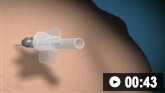 Needle decompression of tension pneumothorax: animated demonstration
How to decompress a tension pneumothorax. Demonstrates insertion of a large-bore intravenous catheter into the fourth intercostal space in an adult.
Cardiac tamponade
Clinical symptoms and signs include chest pain that is relieved on sitting forward, dyspnea, tachycardia, reduced heart sounds, distended jugular veins, and pulsus paradoxus.
Urgent resuscitation (ABC) with supplemental oxygen, and fluid or blood replacement if there is hypovolemia, is required.
Emergency drainage of a pericardial effusion that is causing tamponade is a life-saving procedure.
Pericardiocentesis can be performed without imaging guidance or with the aid of echocardiography or fluoroscopy, with or without hemodynamic monitoring.
Surgical drainage is indicated if there is a hemopericardium, purulent effusion, trauma or associated neoplastic disease.
A chest x-ray should be carried out in order to ensure a pneumothorax has not occurred as a result of the procedure.
Septic shock
Sepsis is a spectrum of disease, where there is a systemic and dysregulated host response to an infection.[36] Presentation ranges from subtle, nonspecific symptoms (e.g., feeling unwell with a normal temperature) to severe symptoms with evidence of multiorgan dysfunction and septic shock. Patients may have signs of tachycardia, tachypnea, hypotension, fever or hypothermia, poor capillary refill, mottled or ashen skin, cyanosis, newly altered mental state or reduced urine output.[37] Sepsis and septic shock are medical emergencies.
Risk factors for sepsis include: age under 1 year, age over 75 years, frailty, impaired immunity (due to illness or drugs), recent surgery or other invasive procedures, any breach of skin integrity (e.g., cuts or burns), intravenous drug misuse, indwelling lines or catheters, and pregnancy or recent pregnancy.[37]
Early recognition of sepsis is essential because early treatment improves outcomes.[37][38][Evidence C][Evidence C] There is an inverse linear correlation between systolic blood pressure and mortality in older people admitted to hospital with suspected infection.[39] However, detection can be challenging because the clinical presentation of sepsis can be subtle and nonspecific. A low threshold for suspecting sepsis is therefore important. The key to early recognition is the systematic identification of any patient who has signs or symptoms suggestive of infection and is at risk of deterioration due to organ dysfunction. Several risk stratification approaches have been proposed. All rely on a structured clinical assessment and recording of the patient’s vital signs.[37][40][41][42][43] It is important to check local guidance for information on which approach your institution recommends. The timeline of ensuing investigations and treatment should be guided by this early assessment.[42]
Treatment guidelines have been produced by the Surviving Sepsis Campaign and remain the most widely accepted standards.[38][44] Recommended treatment of patients with suspected sepsis is:
Measure lactate level, and remeasure lactate if initial lactate is elevated (>18 mg/dL [>2 mmol/L])
Obtain blood cultures before administering antibiotics
Administer broad-spectrum antibiotics (with methicillin-resistant Staphylococcus aureus [MRSA] coverage if there is high risk of MRSA) for adults with possible septic shock or a high likelihood for sepsis
For adults with sepsis or septic shock at high risk of fungal infection, empiric antifungal therapy should be administered
Begin rapid administration of crystalloid fluids for hypotension or lactate level ≥36 mg/dL (≥4 mmol/L). Consult local protocols.
Administer vasopressors peripherally if hypotensive during or after fluid resuscitation to maintain mean arterial pressure ≥65 mmHg, rather than delaying initiation until central venous access is secured. Norepinephrine (noradrenaline) is the vasopressor of choice
For adults with sepsis-induced hypoxemic respiratory failure, high flow nasal oxygen should be given.
Ideally these interventions should all begin in the first hour after sepsis recognition.[44] For adults with possible sepsis without shock, if concern for infection persists, antibiotics should be given within three hours from the time when sepsis was first recognized.[38] For adults with a low likelihood of infection and without shock, antibiotics can be deferred while continuing to closely monitor the patient.[38]
For more information on sepsis, please see Sepsis in adults and Sepsis in children.
Anaphylactic shock
There may be a history of exposure to new foods, drugs, blood transfusions, rashes, bites, or stings.
Features may include: bronchospasm, skin rash, inspiratory stridor, anxiety, nausea, and vomiting.
Intramuscular epinephrine (adrenaline), urgent airway control and supportive therapy should be provided immediately.[30][45][46]
Intramuscular epinephrine can be repeated according to local protocols.
Antihistamines, inhaled beta-adrenergic agents, and intravenous corticosteroids may be considered in cardiac arrest due to anaphylaxis, although there is no evidence that supports the routine use of either steroids or antihistamines in initial resuscitation. Antihistamines and intravenous corticosteroids do not appear to alter the progress of anaphylaxis or prevent biphasic anaphylaxis.[30][46]
All potentially offending agents should be stopped.
Neurogenic hypotension
Should be suspected when hypotension occurs after spinal anesthesia, or in a patient with recent trauma to the spine or brain. It is associated with bradycardia, neurological dysfunction, and the presence of warm, dry skin.
If spinal trauma is suspected, the patient's spine should be immobilized safely, using a spinal board and cervical spine immobilization. Referral to trauma team management is recommended in these patients.
Immediate management priorities are supportive care (including central line placement, volume resuscitation and vasopressors) and imaging of the brain or spinal cord if physical injury is suspected.
Addisonian (adrenal) crisis
The patient may or may not have a known history of adrenal insufficiency. Patients taking chronic glucocorticoids may have secondary adrenal suppression and are also at risk of adrenal crisis if the glucocorticoids are suddenly withdrawn. During times of illness, trauma, or infection, the dose of glucocorticoids needs to be increased.
Intravenous saline should be administered immediately, pending a definitive diagnosis. It is usually necessary to administer 1 liter rapidly, followed by a subsequent 2 to 4 liters over 24 hours.
Urgent administration of glucocorticoid, either with intravenous hydrocortisone or with intravenous dexamethasone sodium phosphate is usually necessary for 1 to 3 days.[47]
If hypoglycemia is present, administration of intravenous glucose or intravenous saline, supplemented with glucose may be necessary.
Once the patient is more stable, the underlying cause of the crises should be investigated further.
Use of this content is subject to our disclaimer