Approach
Diagnosis of oral mucositis (OM) is typically based upon the clinical history and physical exam.[1][2] The most important diagnostic factor is a history of chemotherapy and/or radiation therapy (to the oral cavity) for the treatment of cancer. In certain circumstances, investigations may be warranted to rule out other conditions that may mimic or complicate mucositis.
History and physical exam
A detailed medical history should be elicited from the patient which should include the type of cancer being treated and the therapy they are receiving (chemotherapy and/or radiation therapy), including regimen and intensity. Intensive chemotherapy regimens, radiation therapy to the oral cavity, fractional radiation schedules, and chemoradiation (for oral cancer and in myeloablative regimens for hematopoietic stem cell transplantation [HSCT]) are strongly associated with increased risk for OM.[4][12][13][20]
Symptoms such as mouth pain and intraoral bleeding should be also elicited and patients should be asked about any difficulties in swallowing and maintaining food and fluid intake. The presence and extent of any impairment in oral intake should be ascertained (e.g., can eat and swallow a modified diet; or unable to adequately eat or drink). Patients may also complain of nausea, vomiting, diarrhea, or abdominal pain as a consequence of associated gastrointestinal mucositis.[34] Patients should be assessed for any associated malnutrition, weight loss, and dehydration as a result of reduced oral intake.[35][36]
On examination, oral mucosal changes should be noted. In OM, these range from erythema to patchy or confluent ulceration (often with a superficial pseudomembranous membrane) or, rarely, overt necrosis.[2] Lesions may be unilateral or bilateral, and are typically limited to nonkeratinized oral mucosa. Common sites include the buccal mucosa, lip mucosa, ventral tongue, and soft palate. [Figure caption and citation for the preceding image starts]: Mucositis: buccal mucosaFrom the teaching collection of Rajesh V. Lalla, DDS, PhD, CCRP, DABOM; used with permission [Citation ends].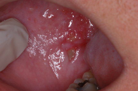 [Figure caption and citation for the preceding image starts]: Mucositis: dorsolateral tongueFrom the teaching collection of Rajesh V. Lalla, DDS, PhD, CCRP, DABOM; used with permission [Citation ends].
In patients receiving standard-dose chemotherapy, lesions typically occur within 2 weeks of initiation, and resolve within 2-4 weeks after treatment is completed.[2] Lesions may last longer in patients receiving high-dose chemotherapy before HSCT; in these patients, resolution of mucositis usually coincides with recovery of neutrophil counts (although this temporal relationship may not be causative).
OM following radiation therapy is limited to the field of radiation. Patients receiving a typical radiation therapy regimen (60 to 70 Gy over 6-7 weeks) for head and neck cancer will usually develop mucosal erythema after 2-3 weeks, followed by ulcerative mucositis over 3-5 weeks.[2] Lesions increase in severity with the dose used, and may take 3-10 weeks to heal following radiation therapy, depending on extent and severity.[1]
The severity of OM can be graded using validated mucositis scales.The World Health Organization scale and National Cancer Institute Common Toxicity Criteria are the two most commonly used criteria.[37][38]
See Diagnostic criteria.
Secondary infection
Frank ulceration of the oral mucosa can act as a portal of entry for host flora to cause local or systemic infection. Therefore, OM lesions may be complicated by secondary viral or fungal infection which can challenge diagnosis.[1][2] Such infection can lead to OM lesions showing delayed healing and occurring at locations unusual for mucositis, such as the keratinized mucosa (hard palate, gingiva, dorsal tongue).[11]
Secondary fungal infection is usually caused by Candida albicans, but other candidal species (e.g., C glabrata, C tropicalis) have also been identified as causative agents.[39] Oral candidiasis is typically not painful. It can manifest as pseudomembranous candidiasis (thrush) or erythematous (atrophic) candidiasis.[Figure caption and citation for the preceding image starts]: Candidal pseudomembranes on the palate, third week of radiation therapy for the management of nasopharyngeal carcinoma. It was diagnosed during the regular, weekly examination. Patient reported xerostomia and mild discomfortFrom the collection of Professor Ourania Nicolatou-Galitis [Citation ends].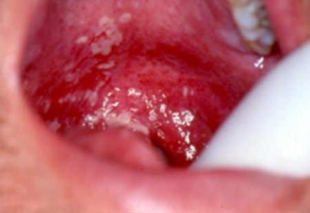
Erythematous candidiasis may present as very red atrophic lesions on the palate, as patchy areas of loss of filiform papillae on the dorsum of the tongue, or as spotted red areas on the buccal mucosa. It cannot be clinically differentiated from the erythematous oral mucosa observed in the early stages of OM during radiation therapy or chemotherapy. A careful history and oral assessment of the patient prior to cancer therapy initiation is helpful in this case.
Secondary fungal infection is common in patients receiving head and neck irradiation and/or chemotherapy, especially those with significant salivary compromise.[2][39][40]
Herpes simplex virus (HSV) infection is associated with vesicular eruption and painful ulceration; however, vesicles rupture quickly, leaving the ulceration visible clinically.[41] Viral infection is more likely in highly immunocompromised patients, such as in those undergoing HSCT.[42][Figure caption and citation for the preceding image starts]: Oral herpes virus infection presenting as painful ulcers covered by pseudomembranes on the right side of the dorsum of the tongue. Diagnosed during the third week of radiation therapy for the management of the maxillary sinus carcinoma. Responded to acyclovirFrom the collection of Professor Ourania Nicolatou-Galitis [Citation ends].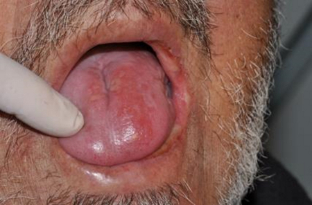
In most cases, mucositis can be differentiated from oral candidiasis and oral herpes infection clinically. Both infections may or may not be superimposed on mucositis.
[Figure caption and citation for the preceding image starts]: Clinical criteria for differential diagnosis of oral mucositis from candidiasis and herpes simplex virus infectionAdapted from Nicolatou-Galitis O et al. The role of benzydamine in prevention and treatment of chemoradiotherapy-induced mucositis. Support Care Cancer. 2021 Mar 1 [Epub ahead of print]. Used under CC BY 4.0 [Citation ends].
Fever may be noted on clinical exam. This may be either related to the presence of secondary infection, or be due to febrile neutropenia following chemotherapy which is an oncologic emergency that requires urgent treatment with empirical antibiotics.
See Febrile neutropenia.
Laboratory investigations
For most patients, no specific investigations are necessary. Where the presentation is suggestive of febrile neutropenia, a complete blood count with differential and blood cultures should be ordered. White blood cell and neutrophil (ANC) counts should be interpreted in the context of any associated malignancy. However, an ANC <500 cells/microliter, or an ANC <1000 cells/microliter with a projected ANC of <500 cells/microliter, along with fever, suggests febrile neutropenia.
If the oral lesions are atypical, or present in unusual locations (e.g., on keratinized mucosa such as the gingiva, hard palate, or dorsal tongue), or persist for longer than expected following the completion of cancer therapy, then additional investigations may be appropriate to rule out alternative or comorbid diagnoses.
Pseudomembranous candidiasis may be confirmed with microscopic examination of superficial smear samples. However, in erythematous candidiasis, hyphae are few in number, and additional testing with culture may be necessary. Viral culture or polymerase chain reaction (PCR) of lesional fluid may confirm HSV infection; PCR has a higher sensitivity than viral culture, but may not be available in some locations.[43]
Use of this content is subject to our disclaimer