Etiology
There are numerous causes of oral ulcerations, and they can be broadly grouped into the following categories:
Traumatic
Metabolic
Dermatologic
Allergic
Immunologic
Infectious
Neoplastic.
However, causes may overlap and, in some people, the etiology is unclear. For example, the commonly observed recurrent aphthous stomatitis (RAS) has been associated with a variety of putative causes including genetic predisposition, stress, trauma, infection, allergy, and nutritional deficiencies.[3][4] None of these etiologies has been validated.
Traumatic causes
Trauma to the oral mucosa may lead to either an immediate or delayed breakdown of the oral mucosa. The injury may be inadvertent or iatrogenic, and the source may be mechanical, thermal, or electrical.[5]
Inadvertent ulcers may occur as a result of:[6]
Simple accidental cheek or tongue biting while chewing or talking
Inappropriate tooth-brushing leading to gingival ulceration
Chemicals/medications (e.g., full-strength hydrogen peroxide applied to the mucosa or aspirin placed in the mouth rather than swallowed)
Thermal burns from drinking or eating beverages or foods that are either too hot or too cold
Natal or neonatal teeth in the newborn (Riga-Fede disease).[7]
Iatrogenic (medical/dental procedure)
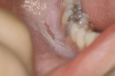
Caused by over manipulation or accidental direct tissue insult. Many cases may not be apparent until after recovery from a local or general anesthetic. Sources to consider include instrumental (e.g., rotary, laser, electrosurgical, blade), chemical (e.g., rinses, topical medications, disinfectants), and physical (e.g., newly placed appliances, stents, brackets) insult.[5][6][Figure caption and citation for the preceding image starts]: Burn from topical aspirinFrom the collection of Dr Huber [Citation ends].
Self-inflicted
Recurrent ulcers caused by self-mutilating conditions may be concurrent with a series of psychiatric disorders and developmental deficiencies or syndromes.[2][8] Examples include genetic disorders (e.g., Lesch-Nyhan, Cornelia de Lange, Tourette, familial dysautonomia, congenital insensitivity to pain, XXXXY syndrome, XXY syndrome, trisomy disorders), psychiatric illness (e.g., Munchausen), encephalitis, coma, bulbar palsy, autism, and intellectual disability.[8] Patient awareness varies from none, as may occur with encephalitis, to compulsive, as may occur with mental illness.
Nutritional causes
Epithelial atrophy, predisposing to ulceration, is a major feature of iron/folate/vitamin B12 deficiency.[9][10]
Iron deficiency can contribute to impaired immune function and epithelial abnormality. It has a female predilection. Iron deficiency in males is uncommon and, if present, suggests occult bleeding (e.g., bleeding ulcer, gastrointestinal malignancy). Patients may have a history of prior anemia, dieting, or alcoholism, and absorptive disorders (e.g., Crohn disease, celiac disease, ulcerative colitis) may be noted. Constitutional signs and symptoms of pallor, fatigue, malaise, shortness of breath, tachycardia, headache, and irritability may be present.[11][12] Chronic ulcerations affecting the tongue (glossitis) are often seen with angular cheilitis and mucosal pallor.
Folate and vitamin B12 are necessary for DNA synthesis. Deficiency of either one or both results in megaloblastic anemia.[9][13] Patients present with chronic nonspecific mucosal ulcerations, a beefy red tongue, and angular cheilitis. Folate deficiency is usually caused by dietary lack, while vitamin B12 deficiency is usually caused by parietal cell antibodies that block B12 absorption (pernicious anemia).[11][13] Correctly identifying the cause of the deficiency is essential before initiating therapy. While folate supplementation may improve the macrocytosis associated with either deficiency, it will not address neurologic pathology associated with pernicious anemia.[12]
Vitamin C is required for normal collagen synthesis and vascular integrity.[14] Patients with vitamin C deficiency may present with gingival edema, bleeding, ulcerations, secondary bacterial infections in the mouth, and loosening of the teeth.[12] In most people, symptoms resolve after vitamin C supplementation.
Dermatologic causes
Oral ulcerations can be a manifestation of several dermatologic diseases. While the concurrent presence of cutaneous lesions often leads to a quick diagnosis, the clinician must always consider that oral ulcerations of a dermatologic disease may occur without cutaneous involvement. These ulcers tend to be chronic, and recognition should lead to appropriate referral and treatment. The more commonly encountered conditions include oral lichen planus, pemphigus vulgaris, and mucous membrane pemphigoid. Less common conditions include chronic ulcerative stomatitis, paraneoplastic pemphigus, epidermolysis bullosa acquisita, lupus erythematosus, and linear IgA bullous dermatosis.[15][16][17] Immunofluorescence is important in diagnosing autoimmune bullous skin disorders.[18]
Lichen planus: a commonly occurring T-cell-mediated dermatologic condition.[19][20][21] Occurrence of oral lesions without concurrent skin lesions is fairly common, affecting about 15% to 35% of patients.[21] There is a distinct female-to-male predilection (2:1). While people of any age may be affected, middle age is the most common. The most commonly affected oral site is the buccal mucosa, followed by the tongue, lips, floor of the mouth, palate, and gingiva. Multiple bilateral and symmetric lesions may manifest as a singular or variable mix of reticular (line, papule, plaques), erosive, or ulcerative lesions. Typical lacey white striations (Wickham striae) are usually present.[20][21] Purely reticular presentations are often asymptomatic and discovered incidentally during routine examination. Erosive and/or ulcerative lesions are typically painful and cause the patient to seek care.[Figure caption and citation for the preceding image starts]: Oral lichen planusFrom the collection of Dr Huber [Citation ends].
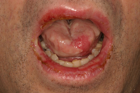
Pemphigus: a group of autoimmune blistering diseases. Pemphigus vulgaris (PV) and paraneoplastic pemphigus can involve the skin and mucosal surfaces of the mouth, eyes, nasopharynx, and esophagus. PV is a rare, potentially life-threatening, intraepithelial splitting disorder with an annual incidence of 1 to 5 cases per million population.[22] It has no sex predilection, and the typical age of onset is 40 to 60 years. Ashkenazi Jews and people of Mediterranean origin have a distinct genetic predisposition for PV.[23] About 90% of patients manifest with chronic oral ulcerations, with areas often subjected to trauma (e.g., buccal mucosa, tongue, palate) being the most commonly affected. Oral lesions are the first manifestation of the disease in >50% of patients.[24][25] The typical oral lesion appears as a painful erosion/ulcer with an irregular border of necrotic epithelium and is partially covered by a fragile membrane.[2] Due to their fragile nature, intact intraoral bullae are extremely unlikely to be seen. The Nikolsky sign tends to be positive (i.e., slight rubbing of the skin exfoliates the outermost layer). Concurrent skin or eye lesions, as well as lesions in the nasopharynx and esophagus, may be present. Two autoantibodies are associated with PV, and their relative amounts determine the clinical phenotype of the disease. Antidesmoglein 1 is associated with cutaneous lesions, and antidesmoglein 3 is associated with mucosal (oral) lesions.[23][26] Paraneoplastic pemphigus is the least common, but most serious, form of pemphigus.[23] Most cases occur in patients who have already developed cancer. Clinical features include acute or chronic mouth sores (flaccid bullae, irregular erosions, ulcerations) commonly located on the gingiva, buccal mucosa, tongue, and palate; ocular symptoms (conjunctivitis, symblephara); and concurrent skin lesions.[Figure caption and citation for the preceding image starts]: Pemphigus vulgaris in a 65-year-old womanFrom the collection of Dr Huber [Citation ends].

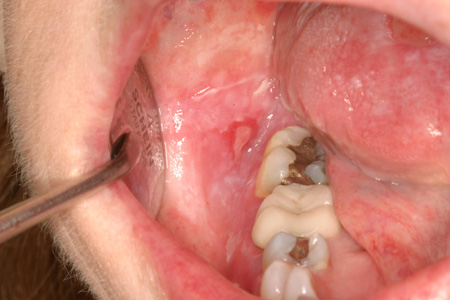
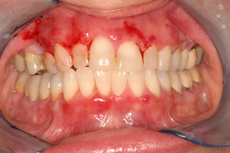
Mucous membrane pemphigoid (MMP): one of a group of uncommon, immune-mediated subepithelial blistering diseases.[2][27][28] It has an annual incidence of 50 to 83 per million population.[28] There is a distinct female-to-male predilection, and the age of onset is 51 to 62 years.[27] MMP primarily involves the oral cavity and conjunctiva.[2] The eyes are involved in up to 40% of patients, which may result in scarring and blindness.[29] The most frequent presentation of MMP in the mouth is desquamative gingivitis (the gingiva has a friable, fiery red, atrophic appearance).[2] The Nikolsky sign tends to be positive.[Figure caption and citation for the preceding image starts]: Mucous membrane pemphigoid in a 53-year-old womanFrom the collection of Dr Huber [Citation ends].
Linear IgA bullous dermatosis: an uncommon, immune-mediated subepithelial blistering disease that usually occurs in children aged <5 years and adults aged >60 years.[30] Pruritic papules, vesicles, and bullae are typically distributed symmetrically over the trunk and extremities.[30] The lesions characteristically resemble a string of pearls (an urticarial plaque surrounded by vesicles).[2] Mucosal involvement is typically found in the oral cavity and conjunctiva.
Epidermolysis bullosa acquisita: an immune-mediated, subepithelial blistering disease with a similar clinical appearance to MMP. Patients have tense blisters and fragile skin, and may have scarring.[2] Progressive and recurrent disease in mucosal tissues can result in irreversible complications including blindness, ankyloglossia (tongue-tie), and esophageal strictures.
Chronic ulcerative stomatitis: a rare, mucocutaneous disorder with predominant oral involvement of the tongue, buccal mucosa, and gingiva.[17][31] It is histologically similar to lichen planus and may have a similar clinical presentation. There is a distinct female-to-male predilection, and it is most common in the fifth and sixth decades of life. Diagnosis of chronic ulcerative stomatitis requires surgical biopsy with immunofluorescence microscopic exam to identify circulating and tissue-bound autoantibodies to a protein, deltaNp63alpha, which is a normal component of stratified epithelia.[17] Patients have a characteristic positive response to hydroxychloroquine therapy.
Allergic/toxic causes
Contact stomatitis: caused by a variety of topical agents including oral hygiene products, foods, and additives.[32] Commonly implicated drugs include barbiturates, lidocaine, gold salts, chlorhexidine, penicillamine, salicylates, and sulfonamides.[33] It typically presents with a single isolated ulcer located on the side of the tongue surrounded by an erythematous halo and is resistant to usual treatments. The prevalence is unknown. Removal of the offending agent resolves the ulcerative lesions.
Oral lichenoid reaction: the number of agents with the potential to cause these ulcerations is extensive and includes dental restorative materials, flavoring agents, and several drugs (the most frequently implicated are nonsteroidal anti-inflammatory drugs [NSAIDs] and ACE inhibitors).[20][21] Drug-related lesions are often mucocutaneous in distribution and resemble lichen planus.[2] Oral lichenoid reaction lesions associated with restorative materials tend to present as an isolated area of involvement near the offending material (e.g., the buccal and/or gingival mucosa in proximity to an amalgam restoration). It may take several months to resolve once the offending agent is removed, which complicates the diagnosis.
Antiresorptive agent-induced osteonecrosis of the jaw (ARONJ): a destructive oral condition manifesting with oral mucosal ulceration and osseous exposure in a patient with past or ongoing exposure to bone-preserving drugs (e.g., bisphosphonate class of drugs and denosumab).[34] Antecedent intraoral trauma (e.g., dentoalveolar surgery) is noted in most patients. Antiresorptive drug uncoupling of the osteoclast-osteoblast balance likely contributes to the aetiopathogenesis of ARONJ. Those at highest risk include oncology patients exposed to prolonged dosing of the most powerful nitrogen-containing bisphosphonate regimens. Patients exposed to low-dose antiresorptive drugs (e.g., osteoporosis prevention) are at much lower risk for ARONJ. Pain is a common presenting complaint. Therapeutic options are limited and focused on eliminating pain, controlling infection, and minimizing further disease progression.[35]
Chemicals/medications: these may include oral rinses, topical medications, or disinfectants.[5][6] Patients undergoing cancer chemotherapy that adversely affects normal proliferation and repair of mucosal tissues may experience ulcerations. Commonly involved medications are: alkylating agents, antimetabolites that affect DNA synthesis, anthracyclines, platinum-based agents, vinca alkaloids, and taxanes.[36][Figure caption and citation for the preceding image starts]: Burn from topical aspirinFrom the collection of Dr Huber [Citation ends].
Erythema multiforme (EM): a group of diseases caused by an allergic reaction to medications, infections, or illness. They are characterized by mucosal erythema and ulcerations with varying degrees of cutaneous involvement.[37][38] The group includes EM minor, EM major, Stevens-Johnson syndrome (SJS), and toxic epidermal necrolysis (TEN).[39] EM major differs from SJS and TEN in that it is associated with frequent recurrences, less fever, milder mucosal lesions, and lack of association with collagen vascular diseases, HIV infection, or cancer.[37] EM typically affects <10% of the body surface area. Its overall prevalence is unknown, as many mild cases likely go undiagnosed.[37] The incidence of TEN is estimated to be 1 to 2 cases per million population per year.[40][Figure caption and citation for the preceding image starts]: Erythema multiforme in a 53-year-old manFrom the collection of Dr Huber [Citation ends].
SJS: a severe form of EM major but is less severe than TEN. As it is associated with significant morbidity and potential death, prompt recognition and management is mandatory.[37][40] Diagnosis is based on the characteristic clinical presentation and skin biopsy results. The incidence of SJS is estimated to be 1 to 6 cases per million population per year.[40]
Immunologic/inflammatory causes
Irregularities in the immune system may underlie the cause of oral ulceration in many patients. Recurrence is common, and the severity can vary from localized, self-limiting trauma to a potentially life-threatening condition. By far the most commonly encountered condition in this category is recurrent aphthous stomatitis (RAS), which is estimated to occur in up to 25% of the population.[3] Other conditions in this category include necrotizing sialometaplasia, Behcet disease, periodic fever syndromes, lupus erythematosus, reactive arthritis, giant cell arteritis, granulomatosis with polyangiitis (formerly Wegener granulomatosis), and graft-versus-host disease (GVHD).
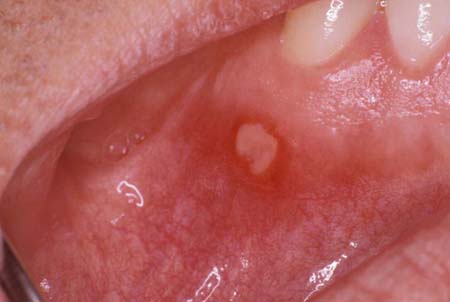
RAS: typically presents in childhood as round or ovoid, shallow ulcerations with a white-yellow pseudomembranous covering.[41][42] RAS has been associated with a variety of putative causes including genetic predisposition, stress, trauma, infection, allergy, and nutritional deficiencies.[3][4] For an otherwise healthy patient, the general rule is that lesions occur only on freely movable mucosa, thus sparing the gingiva and hard palate. Three patterns of presentation are recognized:[42]
Minor RAS: accounts for 80% of cases. Typically, there are 1 to 10 ulcers, <1 cm in diameter, with a characteristic pseudomembranous covering and an intense erythematous margin or halo. Uneventful healing occurs within 14 days.
Major RAS: accounts for 10% to 15% of cases. Ulcers are deeper and may be irregular in shape and >1 cm in diameter. Healing may be prolonged and result in scarring.
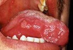
Herpetiform RAS: accounts for 5% to 10% of cases. It is characterized by successive crops of small, 1- to 3-mm round, shallow ulcers. Uneventful healing occurs within 30 days. It is not related to herpes simplex virus (HSV).[Figure caption and citation for the preceding image starts]: Aphthous ulcerFrom the collection of Dr Huber [Citation ends].
 [Figure caption and citation for the preceding image starts]: Major recurrent aphthous stomatitis affecting the soft palateImage created by BMJ Learning [Citation ends].
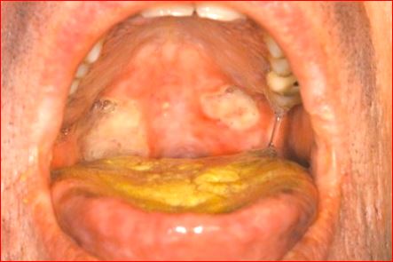
Necrotizing sialometaplasia: a rare disease that produces extensive, deep ulcers with indurated borders, mainly localized to the hard and/or soft palate.[43] It is a benign and self-limiting, non-neoplastic, inflammatory condition of the salivary glands but can simulate a malignant neoplasm. It is believed to be caused by ischemia secondary to trauma or to damage from a chemical or biologic agent.
Behcet disease: a systemic vasculitis classically described as a triad of oral ulcers, uveitis, and genital ulcers.[44][45] Joint symptoms and skin lesions are common, and pathergy (an abnormal reaction to an allergen) is characteristic. It is not clear whether it is an autoimmune or autoinflammatory disease, but there is a strong association with HLA B51.[45] The onset is usually in the third to fourth decades of life, and there may be a family history of the condition.[43] Advanced cases may manifest with macrovascular, central nervous system, and gastrointestinal involvement. Prevalence rates are highest along the historic Silk Road (420/100,000 population in Turkey and 13.5 to 20/100,000 population in the Middle East and Asia).[45] Major, minor, and herpetiform aphthae appear in the oral cavity, generally on the oral mucosa, gingiva, lips, soft palate, and pharynx.[43][Figure caption and citation for the preceding image starts]: Mouth ulcer in Behcet syndromeWith kind permission of Dr Y. Yazici [Citation ends].
Periodic fever syndromes: a constellation of rare autoimmune inflammatory conditions characterized by the presence of aphthous-like ulcerations in association with fever, pharyngitis, and lymphadenopathy.[46] This group includes mouth and genital ulcers with inflamed cartilage (MAGIC) syndrome, periodic fever with aphthous stomatitis, pharyngitis and adenitis (PFAPA) syndrome, and tumor necrosis factor receptor-associated periodic syndrome (TRAPS).
Reactive arthritis (Reiter syndrome): a rare autoimmune condition that develops in response to an infection in another part of the body.[33] Symptoms often include a combination of inflammation of a large joint (e.g., knee), inflammation of the eyes (conjunctivitis and uveitis), and urethritis. In addition, some patients develop mouth ulcers similar to those of RAS. The acute-phase reactants (e.g., erythrocyte sedimentation rate (ESR) or C-reactive protein (CRP)) are often elevated in the acute stage but tend to normalize if the disease becomes chronic. While the presence of HLA-B27 is not specific or required for diagnosis, its presence in the appropriate clinical context improves the accuracy of the diagnosis.
Lupus erythematosus: a chronic inflammatory connective tissue disease associated with the production of multiple autoantibodies, including antinuclear antibodies. Oral lesions occur with skin lesions. They are ulcerated or atrophic, erythematous with a central zone, and surrounded by white, fine, radiating striae.[2] Occasionally, the erythematous atrophic central region may show a fine stippling of white dots. Complications of the lesions include painful ulceration and malignant transformation to squamous cell carcinoma.
Giant cell arteritis: an immune-mediated disease of unknown etiology that is characterized by granulomatous infiltrates within the walls of medium and large arteries of the extracranial branches of the carotid artery.[47][48] It is the commonest of the vasculitides and frequently occurs with polymyalgia rheumatica.[49] Classic signs and symptoms are headache, jaw claudication, scalp tenderness, or acute visual loss. Pertinent laboratory findings include elevated ESR, elevated CRP, and histologic findings of granulomatous infiltration, with or without the presence of giant cells.[50][51] Management includes corticosteroid therapy. While uncommon, involvement of the lingual artery may lead to tongue ischemia and ulceration. More importantly, tongue involvement implies more widespread vessel involvement and the presence of severe disease.
Granulomatosis with polyangiitis (formerly Wegener granulomatosis): an uncommon immune-mediated, necrotizing, granulomatous disease characterized by granulomas of the upper and lower respiratory tracts, generalized vasculitis of small arteries and veins, and glomerulonephritis.[52] Laboratory findings include antineutrophil cytoplasmic antibody, antiproteinase 3 antibody, and antimyeloperoxidase. Oral involvement occurs in up to 13% of patients and may be the first manifestation of the disease. The typical lesion is a hyperplastic petechiae-laden (strawberry) gingivitis. Destructive ulcerative breakdown may occur and extension into the palate is common. Management entails combined corticosteroid and cyclophosphamide therapy.
Chronic GVHD: affects 40% to 70% of engrafted patients. It is the leading cause of morbidity and mortality in hematopoietic stem cell transplantation.[53] Oral features mimic those of lichen planus (lichenoid changes), Sjogren disease (dry mouth), and scleroderma (fibrosis and reduced oral range of motion). Therapy to manage GVHD is intended to diminish pain, allow for eating, improve quality of life, and reduce tissue destruction. The most frequently used agents are cyclosporine and corticosteroids, either alone or in combination.
Infectious causes
Many infectious processes (bacterial, viral, or fungal) can affect the oral mucosa, particularly in an immunocompromised patient. Bacterial conditions include sinus tract infection, necrotizing ulcerative gingivitis, syphilis, gonorrhea, and tuberculosis. Common viral conditions include human herpesviruses 1 (HHV-1) and 2 (HHV-2), varicella zoster virus (VZV), and enteroviruses (herpangina and hand-foot-and-mouth disease). Fungal conditions in this category generally are systemic or locally invasive presentations of zygomycosis, aspergillosis, histoplasmosis, blastomycosis, and paracoccidioidomycosis.[54][55][56] They should be distinguished from the more common oral infection oral candidiasis, which only rarely (e.g., in severe immunosuppression) presents as an ulcerative lesion.
Sinus tract: a chronic infection in the jaw or alveolar process may spread to the surface mucosa, creating a sinus tract (parulis).[57] Patients are often asymptomatic with a history of dental trauma, infection, or untreated dental disease. On examination, there is a sinus tract (parulis), a singular ulcerated or draining exophytic papule (often with evident expressible drainage), and an obvious broken-down tooth or restoration. The maxillary gingiva is the most commonly affected site.
Necrotizing ulcerative gingivitis: an uncommon, painful bacterial infection that typically affects the interdental and marginal gingival tissues in smokers.[58][59] The occurrence of necrotizing ulcerative gingivitis should prompt an investigation of the possibility of an immunocompromised state (e.g., HIV infection). In susceptible patients (e.g., malnourished, immunocompromised), it may progress to the more severe noma (cancrum oris) to involve other orofacial structures.[60][Figure caption and citation for the preceding image starts]: Necrotizing ulcerative gingivitisFrom the collection of Dr Huber [Citation ends].
Syphilis: the oral cavity is the most common extragenital site of syphilis.[2][61] Direct inoculation of the oral mucous membrane from an infected person, usually the result of orogenital or oroanal contact, may result in syphilis.[2][62][63] Most people are easily treated with penicillin.[63] Untreated, syphilis may become more severe.[62] All 3 stages of syphilis may be associated with mucosal ulceration.[62] In primary syphilis, the lesions are known as chancres and occur at the site of penetration of the organism into the mucosa. An oral chancre presents as a solitary, painless, indurated ulceration that persists for 3 to 7 weeks and heals without scarring.[62] The most likely sites of involvement are the lips, tongue, commissures, gingiva, palate, and tonsils.[64] Lymphadenopathy is common.[42] In secondary syphilis, the oral lesions are diverse, including a nonspecific pharyngitis, glistening plaques, and oral ulcers.[62] The most characteristic oral manifestation is the mucous patch. This presents as a shallow, irregularly shaped plaque or ulceration with erythematous borders.[62] A covering gray-white necrotic membrane may be present, and the coalescence of several small patches creates a snail track appearance.[62][65] Lesions of tertiary syphilis manifest as locally destructive granulomas (gummas) or as glossitis with mucosal atrophy. The latter has a tendency for malignant transformation.
Gonorrhea: orogenital contact with an infected person may rarely result in oral inoculation of Neisseria gonorrhoeae.[61][62] Patients can present with multiple nonspecific, fiery red ulcerations with a possible white pseudomembrane.[62] Lymphadenopathy may be present.
Tuberculosis: primary oral tuberculosis is considered rare.[66] Most oral tuberculosis lesions develop secondarily as a consequence of bacteria-laden sputum self-inoculating the oral mucosa. An estimated 0.05% to 5% of patients with active tuberculosis manifest oral lesions.[66] Painful, granulomatous ulceration is typically seen, with possible lymphadenopathy. The lesions may be locally destructive and mimic squamous cell carcinoma.[66] Common sites of occurrence are the tongue, palate, buccal mucosa, and lip.[67]
HHV-1 and HHV-2: these may cause oral ulceration.[68][69][70] About 10% of patients with primary cases have primary herpetic gingivostomatitis, which typically resolves completely within 2 weeks.[69] It can also be a consequence of oral exposure to HSV-2 infection.[69] Recurrent or secondary HSV infection occurs as a consequence of reactivation of latent virus and affects about 20% to 40% of those previously exposed.
VZV: a common HHV.[70] Oral manifestations of primary infection are uncommon but may occur in severe cases.[71] The presence of cropping skin lesions indicate VZV primary infection.[71] Recurrent or secondary VZV infection is known as shingles or zoster and can occur in the oral cavity, particularly in immunocompromised or older people.[72][Figure caption and citation for the preceding image starts]: Primary herpetic gingivostomatitis in a 15-year-old girlFrom the collection of Dr Huber [Citation ends].

Cytomegalovirus: a common HHV. Oral ulceration caused by cytomegalovirus is rare but should be considered in assessing patients who are severely immunocompromised.[73] Coinfection with another herpesvirus such as Epstein-Barr virus or HSV is common.[73][74]
Enteroviruses (herpangina and hand-foot-and-mouth disease): in these conditions, nonspecific oral ulcerations are observed.[75] Uneventful resolution is the usual outcome, but uncommon cases of death due to encephalitis or pulmonary edema have been reported.[76] Herpangina is typically caused by a coxsackievirus group A, and most cases occur in young children, typically <5 years of age. Prodromes include fever, malaise, and headache, and neck or back pain may be present.[75] It usually resolves within 7 days. Hand-foot-and-mouth disease is a highly contagious childhood illness typically caused by coxsackievirus A16 and enterovirus 71. It presents with nonspecific oral ulcerations and characteristic dermal lesions. It usually affects children aged <10 years and resolves within 10 days.[75] There may be a brief prodrome of low-grade fever, malaise, cough, anorexia, abdominal pain, and sore mouth.
Mpox: in the 2022 global mpox outbreak, oropharyngeal/oral lesions have been present in 21% of patients.[77] Oral lesions may be observed in different parts of the oral mucosa including the lips, tongue, and, most commonly, the tonsils.[78][79]
Fungal infections: mycoses associated with oral ulcerations include zygomycosis, aspergillosis, histoplasmosis, blastomycosis, and paracoccidioidomycosis. In healthy patients, ulcerative oral fungal infections are uncommon, but the risk seems increased in conditions such as uncontrolled diabetes mellitus, advanced malignancy (particularly hematologic), or immunosuppression (e.g., HIV).[1][54][55][56] The ulcers often are either an extension of a primary lesion in the paranasal sinus or a manifestation of a systemic mycosis. They typically present as deep-seated ulcers, most commonly affecting the tongue, palate, and maxillary alveolar process.[1][54][55] In zygomycosis, oral ulcerations, sinusitis, or facial cellulitis may be present. In aspergillosis, yellow or black lesions with a necrotic ulcerated base may be present and are typically located on the palate or posterior tongue. In histoplasmosis, chronic nodular, indurated, or granular masses and ulceration with tissue destruction and bone erosion may be observed. Up to 40% to 50% of patients with systemic histoplasmosis manifest oral lesions and the major oral sites affected include the mucosa, tongue, palate, and gingiva. In blastomycosis, single or multiple mucosal ulcerations, sessile projections, and granulomatous or verrucous lesions may be present. Paracoccidioidomycosis is a fungal disease predominantly occurring in Central and South America. Oral lesions are common and typically manifest as oral ulcerative granulomas affecting any part of the oral cavity. Most oral lesions are secondary and arise from inoculation of infected sputum.[56]
Neoplastic causes
Oral cancer ranks as the sixth most common malignancy worldwide and the third most common cancer in developing countries.[80]
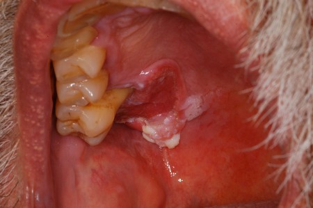
Squamous cell carcinoma: the most common form of oral cancer. Contributing factors include tobacco and alcohol use, HPV infection, immunosuppression, genetic mutations, and diets low in fruit and vegetables.[81][82] Men are affected more frequently than women, and the average age of occurrence is >40 years. Oral carcinoma is typically identified in its later stages, and overall 5-year survival rates do not exceed 60%.[81] Symptoms of more advanced disease include bleeding, loosening of the teeth, difficulty wearing dentures, dysphagia, dysarthria, odynophagia, and development of a neck mass.[83] While any site in the mouth may be affected, the most commonly affected areas include the ventrolateral border of the tongue, the floor of the mouth, and the soft palate complex.[83][84][Figure caption and citation for the preceding image starts]: Oral squamous cell carcinomaFrom the collection of Dr Huber [Citation ends].
Malignant salivary gland tumors (mucoepidermoid carcinoma and adenoid cystic carcinoma): rare and characterized by rapid growth or a sudden growth spurt. They are firm and nodular, and can be fixed to adjacent tissue, often with a poorly defined periphery. Pain and neural involvement are common. Eventually, the overlying skin or mucosa may become ulcerated and the adjacent tissues may be invaded. The most common salivary gland tumors that can present as oral ulceration are mucoepidermoid carcinoma and adenoid cystic carcinoma.[2] Surgery, followed by radiation therapy, is the preferred treatment for resectable disease. There is no effective chemotherapy for salivary gland cancer.
Other oral malignancies: oral melanoma and Kaposi sarcoma may manifest as simple ulcerations.[2] Non-Hodgkin lymphoma may also manifest with oral ulcerations. Oral melanoma is extremely rare, accounting for <1% of primary melanomas. It is characterized by pigmented or amelanotic (white, red, or mucosa-colored) macular lesions of varying size (1 mm to ≥1 cm), predominantly affecting the palate and maxillary gingiva. It is asymptomatic in the early stages of the disease but may lead to loosening of the teeth, bleeding, ulceration, and pain in advanced stages. Oral lesions in Kaposi sarcoma may be the initial site of disease in about 15% of patients with AIDS. Kaposi sarcoma affects the hard palate, gingiva, and dorsum of the tongue and presents as macules, papules, nodules, and exophytic masses of varying size and color. Advanced lesions may become ulcerated from masticatory trauma and secondary infection.
Use of this content is subject to our disclaimer