Approach
Although history and physical examination frequently direct the clinician towards a working diagnosis, ancillary studies are often necessary. Most systemic disorders may be diagnosed with laboratory tests assessing neuroendocrine and ovarian function, and the majority of structural abnormalities are identified through pelvic examination or imaging studies.[1]
History
The mean age of pubertal development aids in deciding the timing of an amenorrhea evaluation. Evaluation should take place with absent menstruation by age 15 years (if other pubertal development is noted), within 3 years of breast development, or with failure of breast development by age 13 years.[1][15][16] Thelarche ("breast budding") denotes previous or current estrogen production. Age of onset or lack thereof may be used to determine when to begin an assessment. Early pubarche (appearance of pubic hair) may be associated with development of polycystic ovary syndrome (PCOS).[17]
Galactorrhea: hyperprolactinemia is more commonly associated with secondary amenorrhea.
History of a traumatic head injury or central nervous system infection: a remote history may be elicited from the patient or parents.
Headache/visual field changes: suggest a central nervous system tumor (e.g., craniopharyngioma).[18]
Anosmia: suggests Kallman syndrome or a complete congenital gonadotropin-releasing hormone (GnRH) deficiency.[19]
Chronic systemic illness: may present with fatigue, malaise, anorexia, and weight loss.
Family history: height should be documented and compared with that of other family members. Short stature is suggestive of Turner syndrome or hypothalamic-pituitary disease.[20] A history of familial delayed puberty, in addition to onset of menarche in the patient's mother and female siblings, should be elicited.
A diagnosis of functional hypothalamic amenorrhea should only be made after excluding anatomic or organic pathology. An inquiry into a patient's health status, eating habits, and body image is necessary. Poor nutritional status due to systemic illness, an eating disorder, and/or low body fat may result in hypothalamic dysfunction.[21] Emotional stress or extreme athleticism can also result in a similar phenomenon.[22][23]
Physical examination
The patient's weight and height should be measured. Shortened height may suggest a chromosomal abnormality. Patients with gonadal dysgenesis and hypoestrogenemia are at risk for shortened final adult height. Growth pattern should be documented and compared with that of first-degree relatives.
On initial examination, careful attention should be given to male pattern baldness, deepening of the voice, wide distribution of terminal hair (male pattern), acne, or oily skin, suggesting hyperandrogenemia. These patterns may vary based on ancestry. If symptoms are slowly progressive, PCOS or non-classic congenital adrenal hyperplasia is possible. If acute and progressive, the patient may be harboring an androgen-producing tumor (ovarian or adrenal).
Consider performing a neurologic examination to assess for neurologic findings such as peripheral vision changes, which suggests an intracranial mass impinging on the optic chiasm (e.g., pituitary adenoma, craniopharyngioma).
Careful examination of the breasts to elicit galactorrhea should be performed in the event that prolactinoma is suspected.
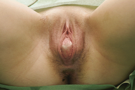
On speculum and bimanual exam, most structural anomalies are identified. The hymen must be assessed first. A blind vaginal pouch will be noted in patients with Mullerian agenesis, transverse vaginal septum, or androgen insensitivity syndrome (the latter along with inguinal hernias). The uterine cervix should be noted on examination. Internal examinations are not always possible and the clinician may need to proceed with imaging options or an examination under anesthesia.[Figure caption and citation for the preceding image starts]: Imperforate hymenLardenoije C, Aardenburg R, Mertens H. Imperforate hymen: a cause of abdominal pain in female adolescents. BMJ Case Reports 2009; doi:10.1136/bcr.08.2008.0722 [Citation ends].
Laboratory tests
All patients with primary amenorrhea, regardless of physical examination findings, should have preliminary laboratory studies drawn, including follicle-stimulating hormone (FSH), estradiol, thyroid-stimulating hormone (TSH), and prolactin. Based on these results, other tests are ordered.
Patients with secondary sexual development should be assessed for pregnancy. A karyotype is indicated for patients with primary amenorrhea and lack of secondary sexual development or those who are diagnosed with premature ovarian insufficiency (usually secondary amenorrhea).[7][24]
FSH: in concert with estradiol levels, gonadotropins help determine if amenorrhea is due to gonadal failure, hypothalamic dysfunction, or systemic or functional causes. FSH is more useful as a single test than luteinizing hormone (LH), and LH is not usually included in the initial investigations ordered.
Serum estradiol: low levels are suggestive of either primary ovarian failure (along with elevated FSH) or suppressed hypothalamic function (low FSH).
Serum prolactin: elevated levels of circulating prolactin (hyperprolactinemia), whether idiopathic or due to a pituitary adenoma, result in hypogonadotropic hypogonadism. For persistently elevated levels, neuroimaging is indicated to rule out intracranial neoplasm.[13]
TSH: is indicated to rule out (primary) hypothyroidism, more commonly associated with secondary amenorrhea. Mild or subclinical hypothyroidism likely will not result in menstrual irregularities.[25] It is proposed that elevated thyrotropin-releasing hormone (TRH) stimulates prolactin secretion from the pituitary, suppressing FSH production.[26]
Serum androgens: done for signs of hyperandrogenism. Androgens such as dehydroepiandrosterone sulfate (DHEAS) and free testosterone will be elevated in patients with PCOS, but higher levels are suggestive of an androgen-producing tumor and these patients should be referred for further investigation.[27][28]
Karyotype: helps to identify patients at risk for gonadal tumors, such as those with premature ovarian insufficiency (usually secondary amenorrhea), androgen insensitivity syndrome, or gonadal dysgenesis.[1][6][14] A diagnosis of complete androgen insensitivity can be confirmed by a 46,XY karyotype, and a diagnosis of Turner syndrome by 45,X. Gonadal dysgenesis (streak gonads) can occur with normal XX and XY karyotypes.[1]
Physiologic tests and imaging
Transabdominal or transvaginal ultrasound is performed if a pelvic examination is not possible. Ultrasound confirms normal anatomy and aids in the diagnosis of most structural abnormalities as well as the presence of an ovarian or adrenal tumor. Transvaginal is the preferred modality, if possible and appropriate, to evaluate endometrial thickness.
MRI is the most effective tool for characterizing specific structural abnormalities and may prevent the need for surgical diagnosis. On MRI, Mullerian agenesis (Mayer-Rokitansky-Kuster-Hauser syndrome) or asymmetrical fusion defects of the Mullerian system (unicornuate uterus) can be identified as well as renal anomalies, which can occur in up to 30% of these patients.[29] A spine x-ray may reveal skeletal abnormalities, which have been reported in around 8% to 32% of patients with Mullerian agenesis.[30]
If prolactin levels are significantly elevated, cranial MRI is indicated to rule out pituitary adenoma.[13]
Bone density measurement may be indicated in selected patients.[24] Bone age is an additional test done for patients with delayed puberty.
Audiometric and ophthalmologic testing is recommended in patients with Turner syndrome. A celiac screen is also useful in these patients.
Use of this content is subject to our disclaimer