Images and videos
Images

Evaluation of paresthesias
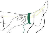
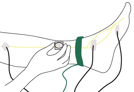
Nerve conduction testing of the lower leg
Created by the BMJ Group
See this image in context in the following section/s:

Evaluation of paresthesias
Central nervous system and non-neurologic causes of paresthesias according to the pattern and level of the lesion
Created by BMJ Knowledge Centre using information from Dr Caroline M. Klein
See this image in context in the following section/s:

Evaluation of paresthesias
Ascending sensory pathways of the spinal cord. The dorsal column system and spinothalamic tract are the major ascending pathways that connect the periphery with the brain.
Betts JG, Young KA, Wise JA et al.Anatomy and physiology. Houston, TX:OpenStax; 2013 (CC BY4.0 - https://creativecommons.org/licenses/by/4.0/)
See this image in context in the following section/s:
Evaluation of paresthesias
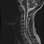
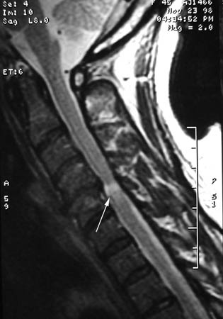
A single level of spinal cord compression with T2 changes, on cervical sagittal T2 sequence in the presence of symptomatic cervical spondylotic myelopathy (CSM)
From the collection of Professor Dennis Turner, Duke University Medical Center; used with permission
See this image in context in the following section/s:
Evaluation of paresthesias
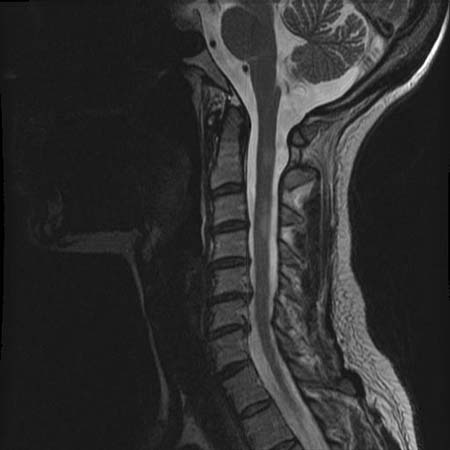
Sagittal T2-weighted MRI showing MS-related myelitis lesion
From the collection of Dean M. Wingerchuk, Mayo Clinic; used with permission
See this image in context in the following section/s:
Evaluation of paresthesias
Peripheral nervous system causes of paresthesias according to the pattern and level of the lesion
Created by BMJ Knowledge Centre using information from Dr Caroline M. Klein
See this image in context in the following section/s:

Evaluation of paresthesias
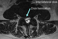
Axial T2-weighted MRI with broad-based lumbar disk herniation predominantly toward the right side
From the collection of Alexios G. Carayannopoulos, Lahey Clinic; used with permission
See this image in context in the following section/s:
Evaluation of paresthesias
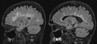
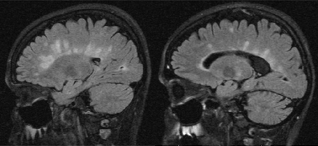
Sagittal FLAIR images with typical MS lesions
From the collection of Lael A. Stone, Cleveland Clinic Foundation; used with permission
See this image in context in the following section/s:
Videos
 Diagnostic lumbar puncture in adults: animated demonstration
How to perform a diagnostic lumbar puncture in adults. Includes a discussion of patient positioning, choice of needle, and measurement of opening and closing pressure.
 Venepuncture and phlebotomy: animated demonstration
How to take a venous blood sample from the antecubital fossa using a vacuum needle.
Use of this content is subject to our disclaimer