The diagnosis of rheumatoid arthritis (RA) is made on the basis of the clinical manifestations of the disease. Laboratory tests or radiographic examinations can be useful in determining prognostic information, but are not essential for making a diagnosis. Patients are referred to a rheumatologist for confirmation of diagnosis.
Classification criteria have been published in an attempt to diagnose RA earlier in the disease course.[48]Aletaha D, Neogi T, Silman AJ, et al. 2010 rheumatoid arthritis classification criteria: an American College of Rheumatology/European League Against Rheumatism collaborative initiative. Arthritis Rheum. 2010 Sep;62(9):2569-81.
https://onlinelibrary.wiley.com/doi/full/10.1002/art.27584
http://www.ncbi.nlm.nih.gov/pubmed/20872595?tool=bestpractice.com
[Figure caption and citation for the preceding image starts]: Rheumatoid arthritis (chronic hand deformities)From the collection of Dr Soumya Chatterjee [Citation ends].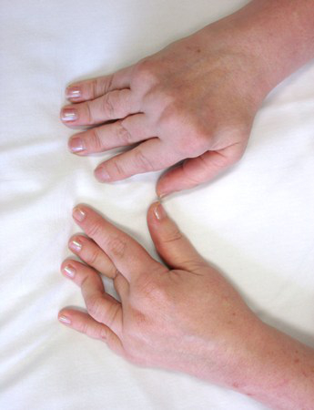
Early diagnosis and treatment is associated with improved outcomes, and is an important principle of management.[49]Fraenkel L, Bathon JM, England BR, et al. 2021 American College of Rheumatology guideline for the treatment of rheumatoid arthritis. Arthritis Rheumatol. 2021 Jul;73(7):1108-23.
https://onlinelibrary.wiley.com/doi/10.1002/art.41752
http://www.ncbi.nlm.nih.gov/pubmed/34101376?tool=bestpractice.com
Workup and treatment should not be delayed while waiting for all RA criteria to be fulfilled; however, there is still a good chance that undifferentiated polyarthritis of <6 weeks' duration will subside spontaneously.
Clinical presentation
Most patients present between the ages of 40 and 60 years.[50]National Rheumatoid Arthritis Society. What is RA? May 2019 [internet publication].
https://nras.org.uk/resource/what-is-ra
Patients who meet diagnostic criteria for RA usually present with a history of bilateral, symmetric pain and swelling of the small joints of the hands and feet that has lasted for more than 6 weeks. Morning stiffness lasting over 1 hour is commonly reported but can also be seen in other inflammatory conditions. Extra-articular features (e.g., rheumatoid nodules over the extensor surfaces of tendons or vasculitic skin involvement) may be seen but are less common.
Swan neck deformity is seen in advanced RA with damage to the ligaments and joints. Classically, there is distal interphalangeal (DIP) hyperflexion with proximal interphalangeal (PIP) hyperextension. Boutonniere deformity is similar, where there is PIP flexion with DIP hyperextension. These deformities are no longer common, as most patients are treated with disease-modifying antirheumatic drugs (DMARDs) at an early stage.
Ulnar deviation, due to inflammation of the metacarpophalangeal (MCP) joints, causes the fingers to become dislocated. As the tendons pull on the dislocated joints, the fingers tend to drift toward the ulnar side.
Extra-articular manifestations seen in more severe disease include pleuritis, interstitial lung disease, pericarditis, and inflammatory eye disease.
Laboratory tests
Once a clinical diagnosis is made, several laboratory tests help to determine prognosis. Rheumatoid factor (RF) is positive in about 60% to 70% of patients with RA.[51]Aho K, Palusuo T, Kurki P. Marker antibodies of rheumatoid arthritis: diagnostic and pathogenetic implications. Semin Arthritis Rheum. 1994 Jun;23(6):379-87.
http://www.ncbi.nlm.nih.gov/pubmed/7524151?tool=bestpractice.com
It is not required for diagnosis but is helpful if present. It should be tested at presentation and does not need to be repeated if positive. The higher the values, the worse the prognosis and the greater the need for aggressive treatment.
Anticyclic citrullinated peptide antibody (anti-CCP), a prognostic marker, is reported in about 70% of patients with RA.[52]Goldbach-Mansky R, Lee J, McCoy A, et al. Rheumatoid arthritis associated autoantibodies in patients with synovitis of recent onset. Arthritis Res. 2000;2(3):236-43.
http://www.ncbi.nlm.nih.gov/pubmed/11056669?tool=bestpractice.com
Anti-CCP can be positive when RF is negative, and it seems to play more of a pathogenic role in the development of RA.[53]van Gaalen FA, Linn-Rasker SP, van Venrooij WJ, et al. Autoantibodies to cyclic citrullinated peptides predict progression to rheumatoid arthritis in patients with undifferentiated arthritis: a prospective cohort study. Arthritis Rheum. 2004 Mar;50(3):709-15.
http://www.ncbi.nlm.nih.gov/pubmed/15022309?tool=bestpractice.com
Anti-CCP does not need to be serially measured, even though it tends to decrease with better disease control.
Erythrocyte sedimentation rate (ESR) or C-reactive protein (CRP) levels are also usually obtained because they reflect the level of inflammation. However, up to 40% of patients with RA may have normal levels.[54]Wolfe F, Michaud K. The clinical and research significance of the erythrocyte sedimentation rate. J Rheumatol. 1994 Jul;21(7):1227-37.
http://www.ncbi.nlm.nih.gov/pubmed/7966063?tool=bestpractice.com
[55]Mohan C, Assassi S. Biomarkers in rheumatic diseases: how can they facilitate diagnosis and assessment of disease activity? BMJ. 2015 Nov 26;351:h5079.
http://www.ncbi.nlm.nih.gov/pubmed/26612523?tool=bestpractice.com
Imaging
Baseline radiographs of the hands and feet are obtained to help with diagnosis and in determining disease severity.[56]Colebatch AN, Edwards CJ, Østergaard M, et al. EULAR recommendations for the use of imaging of the joints in the clinical management of rheumatoid arthritis. Ann Rheum Dis. 2013 Jun;72(6):804-14.
https://ard.bmj.com/content/72/6/804.long
http://www.ncbi.nlm.nih.gov/pubmed/23520036?tool=bestpractice.com
Patients with erosions at baseline who fulfill one of the classification criteria for RA are at risk for severe disease. If imaging the foot and ankle, avoid nonweight-bearing radiographs if the patient is able to stand. This is to ensure the most accurate assessment of the functional bony anatomy.[57]American Orthopaedic Foot & Ankle Society. Five things physicians and patients should question. Choosing Wisely, an initiative of the ABIM Foundation. 2021 [internet publication].
https://web.archive.org/web/20230209030210/https://www.choosingwisely.org/societies/american-orthopaedic-foot-ankle-society
Ultrasound may complement x-ray in the evaluation of suspected RA; it may detect synovitis of the wrist and fingers at the initial presentation.[58]American College of Radiology. ACR appropriateness criteria®: chronic extremity joint pain - suspected inflammatory arthritis. 2022 [internet publication].
https://acsearch.acr.org/docs/3097211/Narrative
http://www.ncbi.nlm.nih.gov/pubmed/28473097?tool=bestpractice.com
[59]Takase-Minegishi K, Horita N, Kobayashi K, et al. Diagnostic test accuracy of ultrasound for synovitis in rheumatoid arthritis: systematic review and meta-analysis. Rheumatology (Oxford). 2018 Jan 1;57(1):49-58.
https://academic.oup.com/rheumatology/article/57/1/49/3061494
http://www.ncbi.nlm.nih.gov/pubmed/28340066?tool=bestpractice.com
Ultrasound may add value in the diagnosis of early seronegative RA.[60]Lage-Hansen PR, Lindegaard H, Chrysidis S, et al. The role of ultrasound in diagnosing rheumatoid arthritis, what do we know? An updated review. Rheumatol Int. 2017 Feb;37(2):179-87.
http://www.ncbi.nlm.nih.gov/pubmed/27803965?tool=bestpractice.com
The presence of erosions, synovial hypertrophy, and hyperemia on ultrasound increases the post-test probability of inflammatory arthritis in seronegative patients.[61]Freeston JE, Wakefield RJ, Conaghan PG, et al. A diagnostic algorithm for persistence of very early inflammatory arthritis: the utility of power Doppler ultrasound when added to conventional assessment tools. Ann Rheum Dis. 2010 Feb;69(2):417-9. [Erratum in: Ann Rheum Dis. 2011 Aug;70(8):1519.]
http://www.ncbi.nlm.nih.gov/pubmed/19359260?tool=bestpractice.com
It is not clear whether the addition of ultrasound to disease activity score strategies is of benefit.[62]Simpson E, Hock E, Stevenson M, et al. What is the added value of ultrasound joint examination for monitoring synovitis in rheumatoid arthritis and can it be used to guide treatment decisions? A systematic review and cost-effectiveness analysis. Health Technol Assess. 2018 Apr;22(20):1-258.
https://www.journalslibrary.nihr.ac.uk/hta/hta22200#/abstract
http://www.ncbi.nlm.nih.gov/pubmed/29712616?tool=bestpractice.com
UK guidelines do not currently recommend ultrasound for routine monitoring of disease activity in adults with RA.[63]National Institute for Health and Care Excellence. Rheumatoid arthritis in adults: management. Oct 2020 [internet publication].
https://www.nice.org.uk/guidance/ng100
Do not order MRI as the initial imaging study to diagnose suspected RA because there is inadequate evidence to justify its use in clinical practice.[64]American College of Rheumatology. Five things physicians and patients should question. Choosing Wisely, an initiative of the ABIM Foundation. 2022 [internet publication].
https://web.archive.org/web/20230209063754/https://www.choosingwisely.org/societies/american-college-of-rheumatology
[58]American College of Radiology. ACR appropriateness criteria®: chronic extremity joint pain - suspected inflammatory arthritis. 2022 [internet publication].
https://acsearch.acr.org/docs/3097211/Narrative
http://www.ncbi.nlm.nih.gov/pubmed/28473097?tool=bestpractice.com
[65]Combe B, Landewe R, Daien C, et al. 2016 update of the EULAR recommendations for the management of early arthritis. Ann Rheum Dis. 2017 Jun;76(6):948-59.
https://ard.bmj.com/content/76/6/948.long
http://www.ncbi.nlm.nih.gov/pubmed/27979873?tool=bestpractice.com
However, MRI can be used as an adjunctive imaging modality when there is diagnostic doubt.[58]American College of Radiology. ACR appropriateness criteria®: chronic extremity joint pain - suspected inflammatory arthritis. 2022 [internet publication].
https://acsearch.acr.org/docs/3097211/Narrative
http://www.ncbi.nlm.nih.gov/pubmed/28473097?tool=bestpractice.com
Disease activity scores
Determining disease activity and presence of poor prognostic factors at diagnosis (functional limitation, extra-articular disease, positive RF, positive anti-CCP, bony erosions on radiograph) should be used to support physician acumen to inform initial treatment decisions.
Composite disease measures are derived from the American College of Rheumatology (ACR) core data set, which includes:
Tender joint count
Swollen joint count
Functional status measured by a health assessment questionnaire (HAQ)
Multidimensional HAQ (MDHAQ) or its derivatives
Pain
Patient and physician global assessment of disease activity, and
Either an ESR or CRP as a marker of inflammation
Any three or more of these combined into a composite index can be used for disease activity monitoring. The most commonly used measures are the disease activity score (DAS), the 28-joint count version of DAS (DAS28), the simplified disease activity index (SDAI), the clinical disease activity index (CDAI), and routine assessment patient index data (RAPID3), all of which are recommended by the ACR.[66]van der Heijde DM, van 't Hof M, van Riel PL, et al. Development of a disease activity score based on judgment in clinical practice by rheumatologists. J Rheumatol. 1993 Mar;20(3):579-81.
http://www.ncbi.nlm.nih.gov/pubmed/8478878?tool=bestpractice.com
[67]Aletaha D, Smolen J. The Simplified Disease Activity Index (SDAI) and the Clinical Disease Activity Index (CDAI): a review of their usefulness and validity in rheumatoid arthritis. Clin Exp Rheumatol. 2005 Sep-Oct;23(5 Suppl 39):S100-8.
http://www.ncbi.nlm.nih.gov/pubmed/16273793?tool=bestpractice.com
[68]Pincus T, Yazici Y, Bergman M, et al. A proposed approach to recognise "near-remission" quantitatively without formal joint counts or laboratory tests: a patient self-report questionnaire routine assessment of patient index data (RAPID) score as a guide to a "continuous quality improvement". Clin Exp Rheumatol. 2006 Nov-Dec;24(6 Suppl 43):S-60-5.
http://www.ncbi.nlm.nih.gov/pubmed/17083765?tool=bestpractice.com
[69]Anderson J, Caplan L, Yazdany J, et al. Rheumatoid arthritis disease activity measures: American College of Rheumatology recommendations for use in clinical practice. Arthritis Care Res (Hoboken). 2012 May;64(5):640-7.
https://onlinelibrary.wiley.com/doi/full/10.1002/acr.21649
http://www.ncbi.nlm.nih.gov/pubmed/22473918?tool=bestpractice.com
Each disease activity measure has its own thresholds of disease activity. For consistency, the same disease activity measure should be used throughout the patient's management. Studies have shown that with close monitoring of disease activity and treating to a target value, it is possible to achieve good responses with any DMARD or combination with biologic agents.[70]Goekoop-Ruiterman YP, de Vries-Bouwstra JK, Allaart CF, et al. Comparison of treatment strategies in early rheumatoid arthritis: a randomized trial. Ann Intern Med. 2007 Mar 20;146(6):406-15.
http://www.ncbi.nlm.nih.gov/pubmed/17371885?tool=bestpractice.com
[71]Grigor C, Capell H, Stirling A, et al. Effect of a treatment strategy of tight control for rheumatoid arthritis (the TICORA study): a single-blind randomised controlled trial. Lancet. 2004 Jul 17-23;364(9430):263-9.
http://www.ncbi.nlm.nih.gov/pubmed/15262104?tool=bestpractice.com
[72]Klarenbeek NB, Güler-Yüksel M, van der Kooij SM, et al. The impact of four dynamic, goal-steered treatment strategies on the 5-year outcomes of rheumatoid arthritis patients in the BeSt study. Ann Rheum Dis. 2011 Jun;70(6):1039-46.
http://www.ncbi.nlm.nih.gov/pubmed/21415052?tool=bestpractice.com