Approach
Unless the underlying cause is already apparent (and being appropriately managed), the presence of pancytopenia always warrants investigation by a hematologist.
The presence of severe pancytopenia (symptomatic anemia [absolute reticulocyte count <60,000/microliter, WBC <500/microliter, and platelets <20 x 10³/microliter]) calls for urgent investigation (within 24-48 hours). A thorough history and physical exam are always required, preferably conducted by a hematologist. A complete blood count and examination of peripheral blood film by a hematologist are essential. Bone marrow exam by aspirate and biopsy is almost always required.[29][30][31]
[Figure caption and citation for the preceding image starts]: Flow diagram for evaluation of pancytopenia. Abbreviations: PNH, paroxysmal nocturnal hemoglobinuria; IBMFS, inherited bone marrow failure syndromesFrom the collection of Jeff K. Davies [Citation ends].
History
The causes of pancytopenia are diverse, and likely causes of pancytopenia differ in children and adults. Particular attention must be paid to patient and family history. Of significance is any history of previous pancytopenia, or single-cell cytopenia, aplastic anemia, inherited bone marrow failure syndromes (IBMFS), early fetal loss, history of cancer, metabolic disorders, liver disease, or connective tissue disorders. A thorough drug history is essential.
The symptoms and signs of pancytopenia relate to the blood cell lineages affected (red blood cells, white blood cells, and platelets). Mild pancytopenia is often symptomless and detected incidentally when a complete blood count is performed for another reason, particularly in association with nonspecific viral illnesses in children. Pancytopenia of this etiology recovers spontaneously.
Spontaneous mucosal bleeding (gums, gastrointestinal tract), petechiae, and purpura with easy bruising secondary to thrombocytopenia are usually the first symptoms to develop directly related to more severe pancytopenia. This is often followed by symptomatic anemia (fatigue, shortness of breath, dependent edema, chest pain in patients with ischemic disease) and bacterial infection secondary to neutropenia (fever, mucositis, abscesses, rigors).
Physical exam
A thorough physical exam is required, preferably by a hematologist. Weight loss and/or anorexia are harbingers of underlying infection (either precedent to the pancytopenia or as a result of it) or malignancy. Spontaneous mucosal bleeding (gums, gastrointestinal tract), petechiae, and purpura with easy bruising secondary to thrombocytopenia are usually the first signs to develop directly related to more severe pancytopenia. These signs are often accompanied by lymphadenopathy (underlying infection, mononucleosis, lymphoproliferative disorder, and malignancy). Abdominal discomfort is a common presentation of splenomegaly and associated conditions. Widespread bone pain and loss of height suggest myeloma, joint pain suggests systemic lupus erythematosus (SLE), and sore throat consideration of mononucleosis.
The following reference points to specific organ systems and associated conditions may be helpful to guide the examination.
Eye examination
Retinal hemorrhage (thrombocytopenia)
Leukemic infiltrates (acute leukemia)
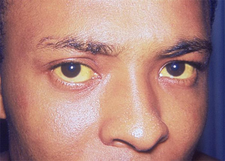
Jaundiced sclera (paroxysmal nocturnal hemoglobinuria [PNH], hepatitis, cirrhosis)
Epiphora (dyskeratosis congenita)[Figure caption and citation for the preceding image starts]: Icterus or jaundiceCDC. Dr Thomas F. Sellers/Emory University; used with permission [Citation ends].
Oral examination
Oral petechiae or hemorrhage (thrombocytopenia)
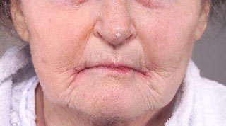
Stomatitis or cheilitis (neutropenia, vitamin B12 deficiency)
Gingival hyperplasia (leukemia)
Oral candidiasis or pharyngeal exudate (neutropenia, herpes family virus infections)[Figure caption and citation for the preceding image starts]: Angular cheilitisFrom the collection of Dr Wanda C. Gonsalves; patient consent obtained [Citation ends].
 [Figure caption and citation for the preceding image starts]: Gingival enlargement, petechiae and bleeding in acute myeloid leukemiaCollection of Giuseppina Campisi, DDS, MS and Giuseppe Pizzo, DDS [Citation ends].
Cardiovascular examination
Tachycardia, edema, congestive cardiac failure (all signs of symptomatic anemia)
Evidence of prior cardiac surgery (cardiac disease associated with congenital syndromes)
Respiratory examination
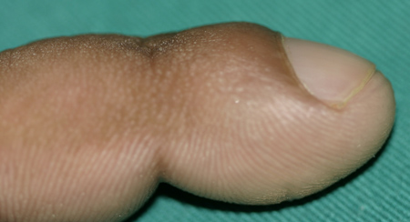
Clubbing (lung cancer)
Tachypnea (sign of symptomatic anemia)[Figure caption and citation for the preceding image starts]: Clubbing of nails showing loss of the classic Lovibond angleFrom the collection of Dr Murlidhar Rajagopalan [Citation ends].
Abdominal examination
Right upper quadrant tenderness (hepatitis)
Lymphadenopathy (infection, lymphoproliferative disorder, HIV disease)
Signs of chronic liver disease
Splenomegaly (infection, myeloproliferative and lymphoproliferative disorders)
Skin examination
Malar rash (SLE)
Purpura/bruising (thrombocytopenia)
Reticular pigmentation, dysplastic nails (dyskeratosis congenita)
Hypopigmented areas
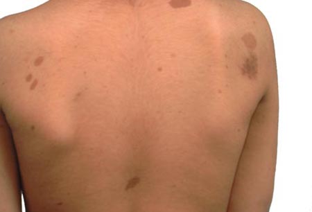
Hyperpigmentation, café au lait (Fanconi anemia)[Figure caption and citation for the preceding image starts]: Café au lait spots on the back of a young boyFrom the personal collection of Dr Vincent M. Riccardi; used with permission [Citation ends].
Musculoskeletal examination
Short stature (Fanconi anemia, other congenital syndromes)
Swelling/synovitis (SLE)
Abnormal thumbs and other radial ray anomalies (e.g., Fanconi anemia)
Signs associated with HIV disease
Morbilliform rash early
Kaposi sarcoma, ulcerating nodules later
Inherited bone marrow failure syndromes (IBMFS) may have characteristic bony, renal, and other congenital abnormalities, or pulmonary or cutaneous abnormalities. A search for these should not be part of an initial workup but, if found on images obtained for other reasons, should prompt further consideration of IBMFS as the etiology of pancytopenia. The absence of physical anomalies does not rule out an IBMFS.
Laboratory
A complete blood count and examination of peripheral blood film by a hematologist are essential. A standard battery of evaluative tests may include:
Serum reticulocyte count
Serum liver function tests and hepatic serology
Serum coagulation profile, bleeding time, fibrinogen, and D-dimer
Serum direct antiglobulin test
Serum B12 and folate
Serum HIV and nucleic acid testing.
Specific testing pinpoints diagnosis in the following conditions:
Fanconi anemia: diepoxybutane (DEB) test for chromosomal breakage in peripheral blood lymphocytes
Lymphoproliferative disorders: immunophenotyping, cytogenetics, lymph node biopsy
Multiple myeloma: immunoelectrophoresis
Paroxysmal nocturnal hemoglobinuria (PNH): peripheral blood immunophenotyping for deficiency of phosphatidylinositol-glycan-linked molecules on peripheral blood cells (e.g., CD16, CD55, CD59)
Cytomegalovirus infection: serum IgM and IgG
Epstein-Barr: serum monospot, viral capsid antigen (VCA), and Epstein-Barr nuclear antibody (EBNA)
Leishmaniasis and other rare infections: blood and bone marrow culture, serum enzyme-linked immunosorbent assay (ELISA)
Rare genetic and metabolic disease: leukocyte glucocerebrosidase activity.
Further specific tests include:
Serum prostate-specific antigen in suspect cases of prostatic malignancy
Telomere length
Genetic analysis involving tests for individual disorders based upon clinical suspicion, or newer panels (many currently in development) to evaluate for mutations and deletions. Germline mutations may be absent in peripheral blood lymphocytes due to mosaicism; consider skin fibroblasts for genetic analysis if there is a strong suspicion of a germline mutation.[32]
Examination of bone marrow is almost always indicated in cases of pancytopenia unless the cause is otherwise apparent (e.g., established liver disease with portal hypertension).[29][30][31] The bone marrow exam consists of both an aspirate and a trephine biopsy, which yield complementary information in this setting. The differential diagnosis of pancytopenia may be broadly classified based on the bone marrow cellularity (reduced cellularity indicates decreased production of blood cells, whereas normal/increased cellularity indicates ineffective production or increased destruction or sequestration of blood cells).
Specifically, bone marrow aspirate permits examination of:
Cytology (megaloblastic change, dysplastic changes, abnormal cell infiltrates, hemophagocytosis, and infection [e.g., Leishman-Donovan bodies])
Immunophenotype (acute and chronic leukemias, lymphoproliferative disorders)
Cytogenetics (myelodysplasia, acute and chronic leukemias, lymphoproliferative disorders).
Bone marrow trephine biopsy permits specific examination of cellularity:
Normal or increased in myelodysplasia, acute and chronic leukemia, myeloma with plasma cells, carcinomatosis marrow infiltration, peripheral destruction/sequestration conditions, early HIV disease, and megaloblastic anemia
Decreased after chemotherapy, acute infection/sepsis, advanced HIV disease, hypoplastic myelodysplastic syndrome, congenital/inherited bone marrow failure syndromes, idiopathic aplastic anemia, systemic lupus erythematosus, and paroxysmal nocturnal hemoglobinuria.
Trephine biopsy also permits examination of histology and evaluation for:
Cellular infiltration
Blasts
Features of myelodysplasia (e.g., abnormal localization of immature precursors)
Reticulin stain (fibrosis).
In the developed world, it has been proposed that the most likely etiology of new-onset pancytopenia, when investigated with bone marrow evaluation, is acute lymphoblastic leukemia in children and acute myeloid leukemia/myelodysplastic syndrome in adults.[33] In some other parts of the world (e.g., India), hypersplenism and infection may be the most frequent etiologies of pancytopenia.[34]

How to take a venous blood sample from the antecubital fossa using a vacuum needle.
Radiology
Abdominal ultrasound scan or computed tomography scan of the abdomen is indicated to evaluate for splenomegaly. Chest radiograph may reveal tumor masses responsible for pancytopenia (e.g., carcinoma, thymoma). In cases where metastatic infiltration of the bone marrow is suspected, thyroid ultrasound or breast imaging may also be appropriate. Inherited bone marrow failure syndromes may have characteristic bony, renal, or pulmonary abnormalities. A search for these should not be part of an initial workup but, if found on images obtained for other reasons, should prompt further consideration of IBMFS as the etiology of pancytopenia.
Use of this content is subject to our disclaimer