Etiology
Infectious gangrene
Necrotizing fasciitis: the most common bacteria isolated are group A beta-hemolytic Streptococcus species, Staphylococcus species, non-group A Streptococcus species, Enterobacteriaceae, and anaerobes such as Clostridium and Bacteroides species.
Gas gangrene: Clostridium perfringens is the most common etiologic agent, with other organisms in this species (C histolyticum, C novyi, C sordellii, C bifermentans, C sporogenes, and C septicum) also being reported.[8] These organisms are spore-bearing, gram-positive, anaerobic bacilli ubiquitous in soil and dust. Nonclostridial organisms have also been isolated in cases of gas gangrene, including Bacteroides fragilis, Escherichia coli, Proteus species, Pseudomonas aeruginosa, and Klebsiella pneumoniae.[8][23]
Ischemic gangrene
Atherosclerosis underlies most peripheral arterial disease, but diabetes-associated microangiopathy is another important factor. Thrombosis associated with hypercoagulable states, intravenous drug abuse, vasculitis, malignancy, or antiphospholipid syndrome and other autoimmune diseases are also important causes.[21] Vasospasm associated with Raynaud phenomenon and cocaine abuse can also give rise to ischemic gangrene.[9]
Inadequate blood supply and gangrene can also result from venous obstruction. Phlegmasia cerulea dolens is a rare condition, usually associated with malignancy, in which there is total or near-total obstruction of venous drainage from a limb.
Pathophysiology
Infectious gangrene
Bacterial multiplication and production of exotoxins require low oxygen tension. The precise role of exotoxins is not entirely clear, although it appears that alpha-toxin is the most important. Alpha-toxin is a metalloenzyme that causes cell destruction by hydrolysis of components of the cell membrane. By this mechanism, it can cause lysis of erythrocytes, leukocytes, platelets, fibroblasts, and muscle cells. It is also thought that this enzyme has C-protein activity.
M proteins have been seen as an important virulence determinant for group A streptococci (GAS). Vimentin on the surface of cells increases binding to GAS at the site of a skeletal injury.
Infection starts with tissue contamination of posttraumatic or postoperative wounds by Clostridium spores. Local wound conditions are more important than the level of clostridial contamination in determining the progression of the condition. Necrotic tissue provides the necessary environment for spore germination; the presence of tissue enzymes and a low oxidation/reduction potential have key roles in this step. Spreading local necrosis of muscle and subcutaneous fat, and thrombosis of blood vessels, create an environment ideal for continued bacterial multiplication. Tissue edema may further compromise blood supply. Fermentation of glucose is probably the main mechanism of gas production.[24]
Ischemic gangrene
Atherosclerosis underlies most peripheral arterial disease. Narrowed blood vessels cannot supply sufficient blood flow to leg muscles and may cause claudication. Atheromatous plaques contain a necrotic core within the arterial intima, consisting of foam cells, cellular debris, and lipids (mainly cholesterol), covered over by a protective cap. Shear forces of the circulating blood or spontaneous rupture can disrupt this protective cap, causing embolization of the cholesterol crystals and inducing thrombogenesis, further reducing blood flow.[21] Critical tissue ischemia is characterized clinically by rest pain, chronic wounds, or tissue necrosis (typically found on the toes).[9] Hypercoagulable states can give rise to large thrombi, which can occlude prominent blood vessels and cause extensive gangrene. Drugs such as cocaine (a powerful sympathomimetic) can cause severe vasospasm sufficient to produce gangrene.[11]
Phlegmasia cerulea dolens is a rare condition, in which there is total or near-total obstruction of venous drainage from a limb. Pain, edema, cyanosis, and ischemia from reduced blood flow ensue, and unless the obstruction is relieved the condition can progress to a form of gangrene known as venous gangrene.
Classification
Infectious gangrene (wet gangrene)
Necrotizing fasciitis: infection of subcutaneous fat and fascia with sparing of skin and underlying muscle[2]
Type I (polymicrobial): caused by Enterobacteriaceae and anaerobes and account for 80% of necrotizing fasciitis.[3] It occurs most frequently after surgical procedures, and in patients with diabetes, alcoholism, immunosuppression, intravenous drug use, or peripheral vascular disease.
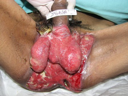
Fournier gangrene is a type I necrotizing fasciitis of the perineal and genital region, resulting from synergistic polymicrobial infection. These are usually from facultative organisms like Escherichia coli, Klebsiella, or enterococci along with anaerobes like Bacteroides, Fusobacterium, Clostridium, or microaerophilic streptococci.[4][Figure caption and citation for the preceding image starts]: Necrotizing fasciitis I subtype involving genital and perineal region; image taken after extensive surgical debridementCollection of Jose Contreras-Ruiz (Clinica del Cuidado Integral de Heridas y Estomas) [Citation ends].
Necrotizing fasciitis of the head and neck is usually caused by mouth anaerobes.
Type II (monomicrobial): rare form of gangrene commonly caused by group A (or C or G) streptococci, which usually develops at a site of trauma on an extremity.[1] It can also be caused by MRSA. Accounts for 20% of necrotizing fasciitis.[3]
Ludwig angina: in the head and neck region, bacterial penetration into the submandibular and sublingual fascial compartments can result in a syndrome known as Ludwig angina, which may develop into necrotizing fasciitis.[5]
Gas gangrene (clostridial myonecrosis) and anaerobic cellulitis (wet necrosis or wet gangrene)
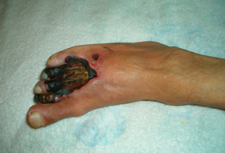
A necrotizing soft-tissue infection with the specific clinical sign of gas formation; it may lead to death. The pathogens most frequently responsible are Clostridium species.[6][7][8] It may occur post-traumatically, postoperatively, or spontaneously. [Figure caption and citation for the preceding image starts]: Eschar and blister formation with notable edema in diabetic patient who developed gas gangrene after a lower limb traumaCollection of Jose Contreras-Ruiz (Clinica del Cuidado Integral de Heridas y Estomas) [Citation ends].

Progressive bacterial (Meleney) synergistic gangrene
This usually occurs after infection at an abdominal operative wound, around an ileostomy or colostomy site, at the exit of a fistulous tract, or in proximity to chronic ulceration on an extremity. It results from a synergistic interaction between Staphylococcus aureus and microaerophilic streptococci.
Gangrenous cellulitis in an immunosuppressed patient[1]
The etiologic considerations for cellulitis occurring in an immunocompromised host include agents that produce such infections in healthy people, as well as a variety of other organisms.
Ischemic gangrene (dry gangrene)
Occurs due to chronic impairment of blood flow. In most patients, the affected part is not infected. This type of gangrene presents clinically as tissue that is cold, black, and dry. In most cases, self-amputation eventually occurs.[9] The pathologic processes involved may include:
Atherosclerosis: associated with peripheral artery disease, diabetes mellitus, smoking[10]
Thrombosis: associated with vasculitis, intravenous drug abuse, trauma, antiphospholipid syndrome, malignancy [Figure caption and citation for the preceding image starts]: Ischemic gangrene secondary to antiphospholipid syndromeCollection of Jose Contreras-Ruiz (Clinica del Cuidado Integral de Heridas y Estomas) [Citation ends].
Vasospasm: associated with Raynaud phenomenon, cocaine abuse.[11]
Although the vast majority of ischemic gangrene arises from arterial compromise, rarely it may also occur due to venous obstruction. This rare condition is known as phlegmasia cerulea dolens and involves total or near-total obstruction of venous drainage from a limb. Pain, edema, cyanosis, and ischemia from reduced blood flow ensue, and unless the obstruction is relieved the condition can progress to a form of gangrene known as venous gangrene.
Alternative classifications
Alternative classification methods exist. Notably, the 2018 World Society of Emergency Surgery consensus guideline divides skin and soft tissue infections (SSTIs) into three main groups - surgical site infections (SSIs), non-necrotizing SSTIs, and necrotizing SSTIs.[12] SSIs are divided into two subgroups – incisional; or organ and organ/space.[13] Incisional SSIs are further divided into superficial (skin and subcutaneous tissue) and deep (deep soft tissue muscle and fascia). Organ and organ/space infections are not true soft-tissue infections. Anatomic extension, purulent or nonpurulent characteristics, and the clinical condition of the patient are proposed as further subgroup variables of necrotizing and non-necrotizing infections.[12]
Use of this content is subject to our disclaimer