Images and videos
Images

Functional neurological and somatic symptom disorders
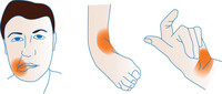
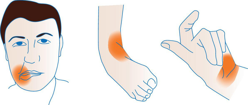
Functional dystonia; orange shading shows typical areas of fixed muscular contraction in functional dystonia
Stone et al. BMJ. 2020 Oct 21;371:m3745; used with permission
See this image in context in the following section/s:
Functional neurological and somatic symptom disorders
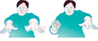
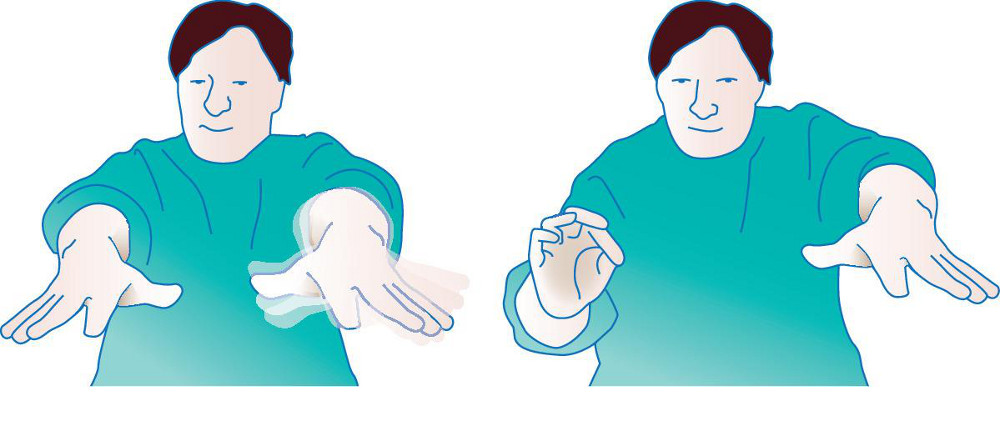
'Entrainment test': watch the other (patient's left) hand while the patient copies the examiner’s rhythmic pincer movements with their right hand; the functional tremor in the left hand stops during the task
Stone et al. BMJ. 2020 Oct 21;371:m3745; used with permission
See this image in context in the following section/s:
Functional neurological and somatic symptom disorders
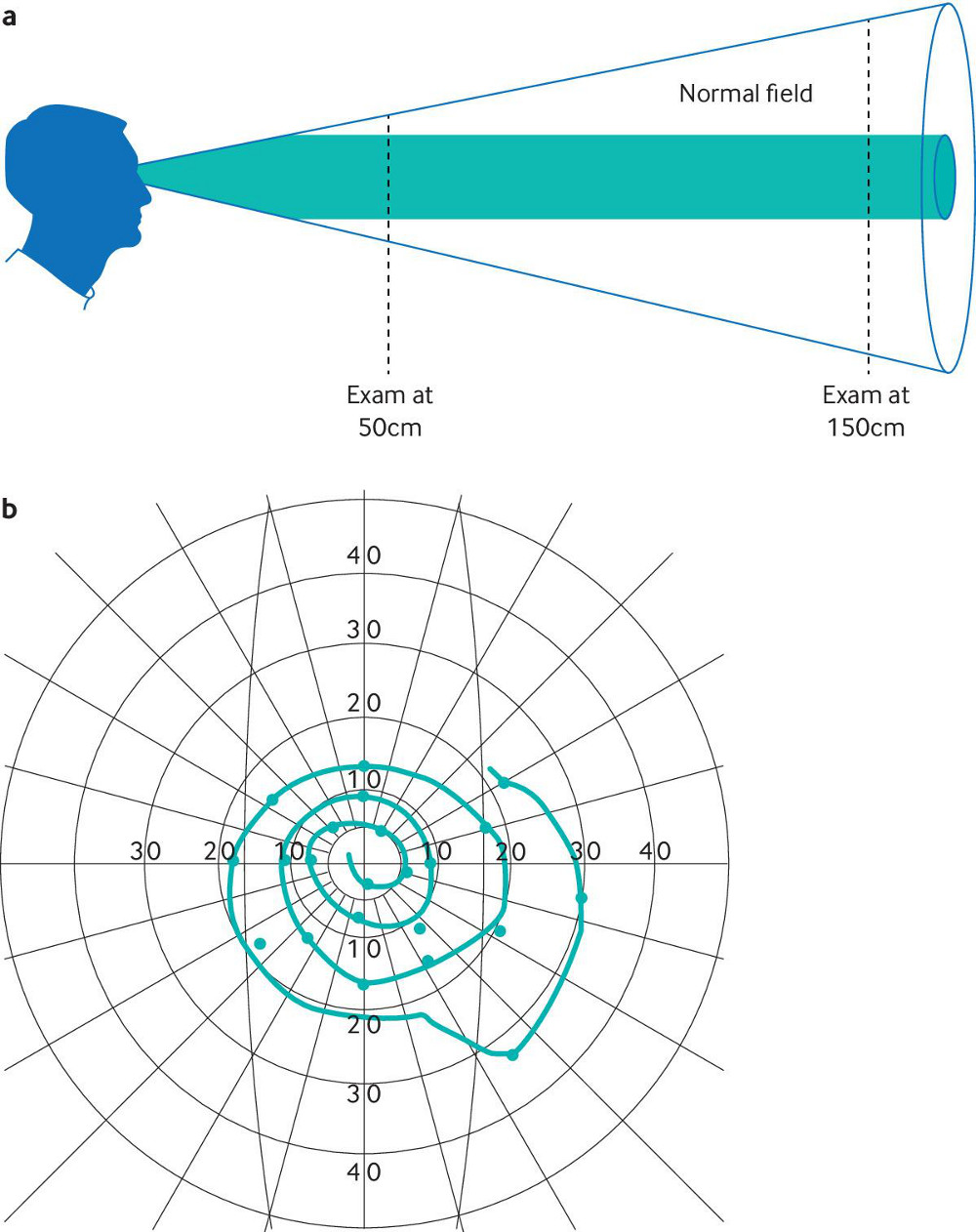
a) A tubular visual field defect at 150cm which is the same width as at 50cm; b) 'Spiralling' on Goldmann perimetry
Stone et al. BMJ. 2020 Oct 21;371:m3745; used with permission
See this image in context in the following section/s:

Functional neurological and somatic symptom disorders
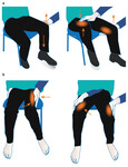
a) Hoover’s sign; hip extension is weak on direct testing (left) but strength becomes normal when there is contralateral hip flexion against resistance; b) Hip abductor sign; hip abduction is weak on direct testing (left) but strength becomes normal when there is contralateral hip abduction against resistance (right)
Stone et al. BMJ. 2020 Oct 21;371:m3745; used with permission
See this image in context in the following section/s:
Use of this content is subject to our disclaimer