Aetiology
Yellow fever virus is an arbovirus of the family Flaviviridae, genus Flavivirus. It is a single-stranded, positive-sense group IV RNA virus that is transmitted by Aedes mosquitoes (mainly Aedes aegypti) in Africa and Aedes andHaemagogus mosquitoes in the Americas. One serotype and at least 5 genotypes have been recognised. It has a viral envelope consisting of a lipid bilayer with dimers of an envelope protein (E) that are involved in the attachment to glycosaminoglycan receptors in the cell wall and internalisation of the virus. Antibodies directed against certain E-protein epitopes disrupt viral internalisation and are considered protective.[2][Figure caption and citation for the preceding image starts]: A female Aedes aegypti mosquitoCDC/Prof. Frank Hadley Collins, University of Notre Dame [Citation ends].
There are three well-documented transmission cycles.
Sylvatic form: maintained within monkey populations in the rain forest, with sporadic transmission to people in close contact with the forest.
Intermediate form: maintained by people and monkeys in rural villages, where increased human transmission leads to small outbreaks.
Urban form: maintained in high-density urban environments and involves domestic mosquitoes, resulting in larger numbers of patients and higher mortality.
[Figure caption and citation for the preceding image starts]: Types of transmission of yellow feverWHO [Citation ends].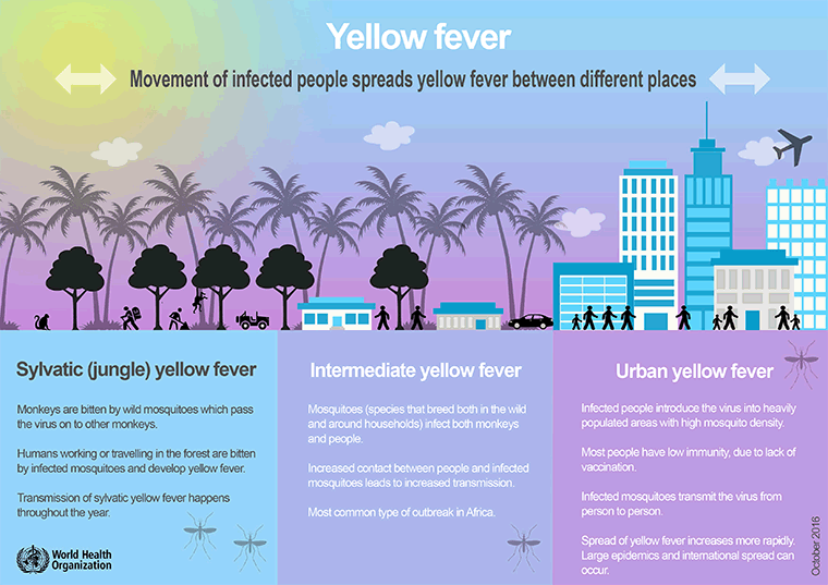
Pathophysiology
The incubation period is 3 to 6 days. After inoculation by an appropriate vector, E proteins facilitate attachment to glycosaminoglycan receptors in the cell wall and internalisation of the virus. The virus initially replicates in lymphatic tissues, followed by viraemia and subsequent dissemination to other organs: in particular, the liver and kidneys. The affected hepatocytes undergo eosinophilic degeneration with condensed nuclear chromatin (Councilman bodies). Midzonal distribution and sparing of cells around the portal tracts and central vein is characteristic.[13] Renal involvement is characterised by eosinophilic changes of the tubular epithelium and fatty changes. The characteristic yellow fever albuminuria generally reflects changes in glomerular function.
Death is preceded by cytokine dysregulation with subsequent cardiovascular shock and multi-organ failure.[14] Interestingly, it is preceded by pseudo-viral clearance during the period of intoxication, when early neutralising antibodies have been generated.[2]
[Figure caption and citation for the preceding image starts]: Yellow fever virus particles viewed through a transmission electron microscopeCDC Public Health Image Library [Citation ends].
Use of this content is subject to our disclaimer