Approach
History and physical examination are critical in establishing the diagnosis and determining the underlying aetiology. Initial work-up involves laboratory tests and a computed tomography (CT) scan of the abdomen and pelvis. A multidisciplinary team including gastroenterologist, surgeon, and critical care specialist should be involved based on a high index of suspicion.
History
The history should focus on the presence of a personal history of inflammatory bowel disease (IBD) or suspicion for infectious agents. Recent use of antibiotics and other medications such as opiate pain medications, anticholinergics, antidepressants, chemotherapy, or antimotility agents can be contributory. Presence of immunocompromised state (e.g., AIDS) and post-transplantation status should be noted.
The majority of patients with toxic megacolon present during an ongoing episode of severe colitis. Therefore, the symptoms associated with colitis, including diarrhoea (watery or bloody), fevers, chills, and crampy abdominal pain, are frequently present before the onset of toxic megacolon. Patients often exhibit typical symptoms of acute colitis for the week or 2 preceding the onset of acute presentation.
Physical examination
Physical examination should begin with assessment of vital signs such as heart rate, blood pressure, and tissue perfusion (urine output). Abdominal examination reveals signs of colitis such as abdominal distension, decreased bowel sounds, tenderness, and signs of peritonitis (focal or diffuse). Systemic manifestations including fever, tachycardia, hypotension, and mental status changes are often present. However, the use of high-dose corticosteroids and analgesics may mask signs of toxic megacolon.[7] Some patients with Clostridium difficile colitis presenting with toxic megacolon may not have a typical history (such as recent antibiotic use), or typical symptoms and signs (such as diarrhoea), making diagnosis more difficult.
Laboratory
Laboratory studies including a full blood count with differential, basic metabolic panel (electrolytes and serum creatinine), serum albumin, and serum lactic acid are mandatory. Blood should be taken for blood cultures at presentation, before the administration of antibiotics if possible. These laboratory studies should be repeated if necessary at least every 24 hours or at shorter intervals, depending on the clinical scenario. Baseline blood gas analysis is sometimes helpful. Stool sample should be sent to test for bacterial and viral infections. C difficile infection should be assessed with a nucleic acid amplification test; repeat testing is not recommended within 7 days.[24]
Patients demonstrate evidence of systemic manifestations including an elevated WBC count with left shift, and elevated CRP and erythrocyte sedimentation rate, though these are not diagnostic of toxic colitis/toxic megacolon. Patients may also present with anaemia due to frequent bloody bowel movements and chronic disease. Electrolyte abnormalities including hypokalaemia are observed due to an inability of the inflamed colon to reabsorb water and salt, and an increased excretion of potassium due to large amounts of diarrhoea.
Imaging
A CT scan of the abdomen and pelvis is diagnostic and often mandatory in the assessment of severity of disease as well as in planning treatment. Besides colonic distension, features of colitis such as mucosal hyperaemia, mesenteric oedema and stranding, and free fluid in the peritoneal cavity can be seen.[25] Any complications of toxic megacolon, such as perforation of the colon (evidenced by free air in the peritoneal cavity or colonic necrosis on the CT scan of the abdomen and pelvis), indicates urgent surgical treatment.
Abdominal x-ray will reveal the extent of colonic dilation and any free air under the diaphragm.[7] An abdominal x-ray can be performed in the accident and emergency department and should be done in unstable patients who cannot be transported safely to the CT scanner. An upright chest x-ray can be helpful in a similar fashion. Diagnostic features on abdominal x-ray include colonic dilation >6 cm, disturbed/absent colonic haustration, air-fluid levels, and small bowel distension.[7] Abdominal plain films are useful for monitoring and should be obtained on admission, and at least daily thereafter until resolution or operative intervention.[26][Figure caption and citation for the preceding image starts]: Abdominal x-ray demonstrating colonic dilationUniversity of Chicago Medical Center; used with permission [Citation ends].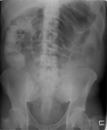
Sigmoidoscopy and biopsy
Flexible sigmoidoscopy will aid in the diagnosis of certain patients, and it may help to identify pseudomembranous colitis, show features of IBD, or identify full-thickness necrosis of the colon. Full sigmoidoscopy is contraindicated due to the risk of perforation; therefore, if performed, the scope should be passed to only 20 cm and with minimal insufflation (CO₂ preferred to air). Biopsy of mucosa and stool samples are often obtained. The presence of peritonitis and severe abdominal distension with tenderness are contraindications to endoscopic evaluation.
Colonoscopy is rarely indicated in this setting.
Use of this content is subject to our disclaimer