History and exam
Key diagnostic factors
common
presence of risk factors
Key factors include genetic predisposition, race, and age.
presence since birth
Congenital nevi are usually present at birth or shortly thereafter.
asymmetrical, indistinct or irregularly bordered, variably coloured papules with diameter >5 mm
Atypical, dysplastic, or Clark's nevi may have a 'fried egg' appearance, with a central papular component and a flat peripheral component, and may have indistinct borders.[30][32]
Larger than 5 mm, and may have variable coloration from pink to tan to brown.
Distinction from a melanoma may be difficult, as many of these criteria are fulfilled by melanoma, but to a greater degree than in dysplastic nevi. [Figure caption and citation for the preceding image starts]: A dysplastic or Clark's nevusFrom the collection of Jason Lee, Thomas Jefferson University [Citation ends].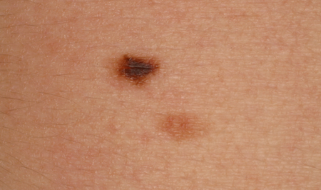 [Figure caption and citation for the preceding image starts]: A dysplastic or Clark's nevusFrom the collection of Jason Lee, Thomas Jefferson University [Citation ends].
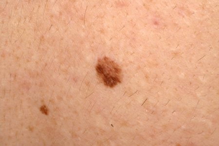 [Figure caption and citation for the preceding image starts]: A dysplastic or Clark's nevusFrom the collection of Jason Lee, Thomas Jefferson University [Citation ends].
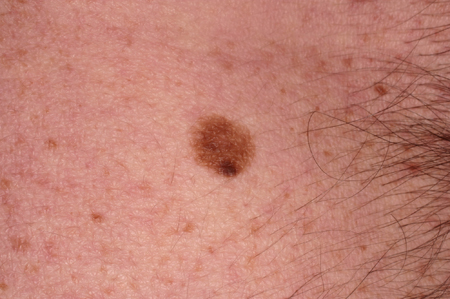
history of change in shape and colour
Melanocytic nevi may evolve and become more raised and dome-shaped over time. They may get lighter or darker over time. Spitz nevi often have a history of rapid growth initially.
asymptomatic (usually)
Nevi are usually asymptomatic, although they may also be reported to be itchy and may become irritated with trauma.
multiple lesions
People who have nevi can have solitary lesions, but often there are multiple lesions.
flat, brown macule
Junctional acquired nevi are often small, flat, uniformly brown macules.
dome-shaped papule
Compound and dermal nevi are often dome-shaped and tend to have uniform pink, tan, or brown coloration. [Figure caption and citation for the preceding image starts]: A common acquired nevusFrom the collection of Jason Lee, Thomas Jefferson University [Citation ends]. [Figure caption and citation for the preceding image starts]: A compound acquired melanocytic nevusFrom the collection of Laurel Schwartz, Thomas Jefferson University [Citation ends].
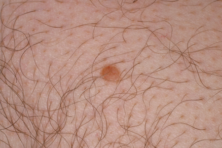
light brown background with speckled darker brown spots within
A nevus spilus has a light brown background with a few scattered darker brown papules or macules within the lesion.[7][Figure caption and citation for the preceding image starts]: A nevus spilusFrom the collection of Jason Lee, Thomas Jefferson University [Citation ends].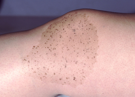
blue-grey dome-shaped papule
Common blue nevi are solitary blue-grey to black papules or flat macules that are usually <1 cm and often found on the dorsal extremities, head and neck, and sacrum.[7][Figure caption and citation for the preceding image starts]: A blue nevusFrom the collection of Jason Lee, Thomas Jefferson University [Citation ends].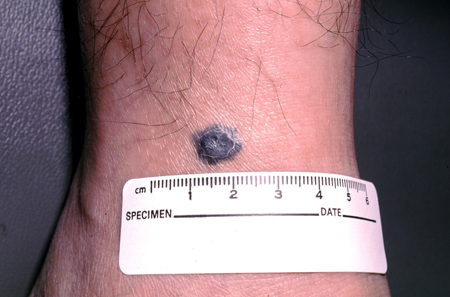 [Figure caption and citation for the preceding image starts]: A blue nevusFrom the collection of Jason Lee, Thomas Jefferson University [Citation ends].
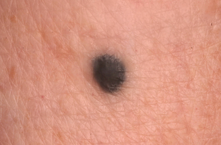
central pink-to-brown papule with a surrounding depigmented white ring
The term 'halo nevus' describes a morphological feature of an inflamed nevus, thought to be part of an immune response to nevus antigens.[7]
Halo nevus usually heralds the disappearance of the nevus, leaving a white, depigmented macule, which may eventually repigment. [Figure caption and citation for the preceding image starts]: A halo nevusFrom the collection of Jason Lee, Thomas Jefferson University [Citation ends].
pinkish-brown papule
Spitz nevi are often found on the face and lower extremities as pink or red-brown papules with a smooth or verrucous surface.[7][Figure caption and citation for the preceding image starts]: A Spitz nevus on the earFrom the collection of Jason Lee, Thomas Jefferson University [Citation ends].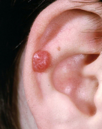
Risk factors
strong
genetic predisposition
Use of this content is subject to our disclaimer