Aetiology
Factors including genetics, immunology, and infection may contribute.
Genetics
Psoriasis heritability is between 60% to 90%, which is higher than most other multifactorial diseases.[10] Studies of monozygotic twins, linkage studies, and genome-wide association studies provide evidence that psoriasis has a genetic predisposition.[11][12][13]
Most genes associated with psoriasis are involved in the immune response, and relatively few encode for skin-specific proteins. HLA-Cw6 encodes an antigen involved in T-cell activation. There is an increased prevalence of HLA-Cw6 in people with psoriasis compared with controls. Tumour necrosis factor (TNF)-alpha, another protein-coding gene associated with innate and adaptive immune response, is implicated in the aetiology of psoriasis. Pathogenic involvement of genes related to Th17-cell activation have been demonstrated in people with psoriasis.[12][13][14][15][16]
Immunology
Psoriasis is believed to be triggered by external insults in genetically susceptible individuals. Recognised triggers include trauma, infection, and medications (e.g., beta-blockers, lithium). Following initiation by an insult, the host DNA forms complexes with antimicrobial peptides released from keratinocytes (skin cells), which results in inflammation and keratinocyte proliferation to cause disease presentation.[16][17][18]
Infection
Guttate psoriasis is often observed subsequent to upper respiratory infection, such as streptococcal pharyngitis. It may also be associated with HIV infection. Viral infection, immunisation, and any intercurrent illness have been linked to flares of guttate and plaque psoriasis.[16][19]
Pathophysiology
Psoriasis is a hyperproliferative disorder, involving a complex cascade of inflammatory mediators. Mitotic activity of basal and suprabasal cells is significantly increased, with cells migrating from the basal layer to the stratum corneum in just a few days. The silver scale on the surface of psoriasiform lesions is simply a layer of dead cells.[16][20]
Early clinical studies of TNF inhibitors demonstrated the important role of these cytokines in psoriasis, prompting the condition to be regarded as primarily driven by T-helper-1 (Th-1) cells.[21] However, evidence supports the pivotal involvement of a different immunological axis underlying the pathogenesis of psoriasis; namely, T-helper cells producing interleukin (IL)-17 and IL-23.[22][23][24] IL-17 and IL-23 expression is increased in the serum, lesional skin, uninvolved skin, and even tear liquid of patients with psoriasis compared with patients without psoriasis. These cytokines are now considered to be central to the pathogenesis of psoriasis as demonstrated by the efficacy of therapeutics inhibiting IL-17 or IL-23 pathways.
The presence of T-cells reacting against autoantigens has been detected; three autoantigens have been identified: LL37, ADAMTS-like protein 5, and phospholipase-2-derived products.[25][26][27][28]
Classification
International Psoriasis Council[2]
1. Plaque psoriasis
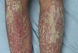
Raised inflamed plaque lesions with a superficial silvery-white scaly eruption. The scale may be scraped away to reveal inflamed and sometimes friable skin beneath.[Figure caption and citation for the preceding image starts]: Plaque psoriasis on legsFrom the collection of Professor Tsu-Yi Chuang, MD, MPH, FAAD; used with permission [Citation ends].
 [Figure caption and citation for the preceding image starts]: Plaque psoriasis on backFrom the collection of Professor Tsu-Yi Chuang, MD, MPH, FAAD; used with permission [Citation ends].
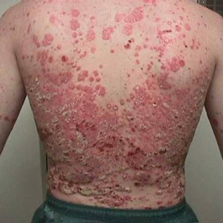 [Figure caption and citation for the preceding image starts]: Plaque psoriasis on kneeFrom the collection of Professor Tsu-Yi Chuang, MD, MPH, FAAD; used with permission [Citation ends].
 [Figure caption and citation for the preceding image starts]: Plaque psoriasis on footFrom the collection of Professor Tsu-Yi Chuang, MD, MPH, FAAD; used with permission [Citation ends].
 [Figure caption and citation for the preceding image starts]: Plaque psoriasis on scalpFrom the collection of Professor Tsu-Yi Chuang, MD, MPH, FAAD; used with permission [Citation ends].
2. Guttate psoriasis
Widespread, erythematous, fine, scaly papules (water drop appearance) on trunk, arms, and legs. The lesions often erupt after an upper respiratory infection.[Figure caption and citation for the preceding image starts]: Guttate psoriasisFrom the collection of Professor Tsu-Yi Chuang, MD, MPH, FAAD; used with permission [Citation ends].

3. Pustular psoriasis
Acute generalised pustular psoriasis (von Zumbusch): rare, severe, urgent.
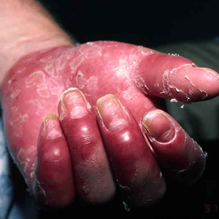
Palmoplantar pustulosis: chronic involvement of hands and feet.[Figure caption and citation for the preceding image starts]: Pustular psoriasisFrom the collection of Professor Tsu-Yi Chuang, MD, MPH, FAAD; used with permission [Citation ends].

4. Erythroderma (erythrodermic psoriasis)
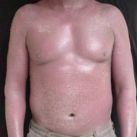
Generalised erythema with fine scaling. It is often associated with pain, irritation, and sometimes severe itching.[Figure caption and citation for the preceding image starts]: ErythrodermaFrom the collection of Professor Tsu-Yi Chuang, MD, MPH, FAAD; used with permission [Citation ends].
5. Psoriatic arthritis
Unique in that, in addition to skin lesions, there is joint involvement that causes inflammatory damage and deformity. It affects approximately 20% of people with psoriasis, as reported by a 2019 epidemiological study.[3]
Most people with nail psoriasis have psoriatic arthritis. The arthritis most commonly involves fingers, hands, toes, and feet, and, less commonly, knees, elbows, and axial and sacroiliac joints.
Cutaneous psoriatic lesions precede arthritis in 70% of cases.
Psoriatic arthritis causes stiffness, inflammation, pain, and progressive and permanent joint damage. The arthritis is asymmetrical in around 50% of cases.[Figure caption and citation for the preceding image starts]: Psoriatic arthritisFrom the collection of Professor Tsu-Yi Chuang, MD, MPH, FAAD; used with permission [Citation ends].
 [Figure caption and citation for the preceding image starts]: Nail psoriasis - pitted nailsFrom the collection of Professor Tsu-Yi Chuang, MD, MPH, FAAD; used with permission [Citation ends].
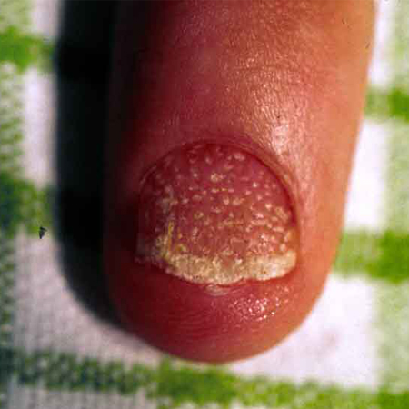
6. Keratoderma blennorrhagicum (reactive arthritis)
Reactive immune disorder characterised by psoriasiform plaques, urethritis/cervicitis, conjunctivitis, and arthritis.
Circular, scaly, scalloped-edged hyperkeratotic psoriasiform papules and plaques, which is sometimes painful and pustular (at the centre of lesions), appears on soles and toes, and, less commonly, legs, palms, scalp, and penis.
Occurs in genetically susceptible people with HLA-B27 following an infection (particularly Yersinia enterocolitica and Yersinia pseudotuberculosis).
Use of this content is subject to our disclaimer