Approach
History and physical examination are critical components of the evaluation of the febrile child and frequently result in identifying the underlying cause of the fever.[2][11]
History
Fever
The characteristics of the fever are important, including height and duration of fever and method of temperature measurement. The subjective determination of fever at home by a carer is reasonably accurate.[29][30] The incidence of serious bacterial infection may be higher in children with higher temperatures[31][32] and a shorter duration of symptoms. Night-time fever with sweats may suggest tuberculosis or a lymphoma.
The response to antipyretic therapy does not differentiate between viral and bacterial causes.[33]
Low-grade prolonged fever may be more common with viral infections, inflammatory/vasculitic disorders, and malignancies.
Rash
Fevers in children are sometimes accompanied by rashes. Examination of the characteristics and distribution of the rash may help in determining the cause of fever.
A maculopapular, petechial, or urticarial rash may be present with bacterial infections or with medication-related fever.[34]
Viral infections may present with a non-specific maculopapular or a petechial rash.[34]
Palpable purpura suggest vasculitis, rheumatological causes, drug reactions, infections, and malignancy.
Erythema migrans, usually at the site of a tick bite, may be present in Lyme disease.[35]
Accompanying symptomatology
Inflammatory/vasculitic disorders and malignancies can result in multisystem involvement such as:
Pleuritis, carditis, or pericarditis resulting in symptoms of cough and breathlessness
Gastrointestinal symptoms of nausea, vomiting, diarrhoea, and abdominal pain
Central nervous system (CNS) symptoms of confusion, seizures, altered mental state, and focal neurological deficits
Renal involvement with oedema and pruritus
Musculoskeletal involvement resulting in arthralgia, myalgia, and bone pain.
Arthralgias may also be present with bacterial infections such as septic arthritis and osteomyelitis; viral infections such as infectious mononucleosis, cytomegalovirus (CMV), toxoplasmosis, and tick-borne illnesses (Lyme disease, Rocky Mountain spotted fever, or dengue fever); medication-related reactions; or malignancies.
Bone pains are present in osteomyelitis, leukaemia, juvenile idiopathic arthritis (JIA), and systemic lupus erythematosus (SLE).
Cardiorespiratory symptoms may be observed with bacterial infections such as pneumonia or endocarditis, viral infections (including myocarditis), tuberculosis, thyroid storm, and inflammatory/vasculitic disorders. Persistent nasal discharge is associated with sinusitis.
ENT symptoms such as pharyngitis, otalgia and persistent nasal discharge may accompany ENT infections, such as sinusitis, acute otitis media and tonsillitis.
Gastrointestinal symptoms such as nausea, vomiting, abdominal pain, and diarrhoea are associated with myocarditis, inflammatory bowel disease (Crohn's disease, ulcerative colitis), other inflammatory/vasculitic disorders, typhoid fever, hepatic abscess, medication-related reactions, heat-related illness, autonomic disorders, and thyroid storm.
CNS symptoms such as confusion, altered mentation, seizures, and focal deficits may be observed with encephalitis, bacterial endocarditis, myocarditis, cerebral abscesses, tuberculous meningitis, secondary spread from a malignancy, severe inflammatory disorders (e.g., SLE), and cerebral malaria. Chorea is present with advanced rheumatic fever.
Urinary symptoms of dysuria, frequency, and haematuria accompany urinary tract infections (UTIs) such as cystitis and pyelonephritis.
Poor growth, poor appetite, and weight loss are observed with any chronic illness as opposed to a history that is of relatively short duration (e.g., bacterial infections).
Absence of tears can be observed in autonomic disease.
There may be a history of recent tick or mosquito bite with Lyme disease, Rocky Mountain spotted fever, malaria infection, and dengue fever.
Medication and immunisation history
A critical component of the history is the child's immunisation and medication history. Unimmunised or under-immunised patients are at higher risk for certain infections (e.g., Streptococcus pneumoniae, Haemophilus influenzae type b). Recent history of vaccines may point towards a vaccine reaction; exposure to serum products may indicate a serum sickness reaction. Although a child who has received at least two doses of a pneumococcal conjugate vaccine is at substantially lower risk for invasive pneumococcal disease, Streptococcus pneumoniae remains a leading cause of serious bacterial infection in children.[36]
Ingestion of various drugs such as anticholinergic medications, amphetamines, and cocaine can result in drug-related fever. Many medications are also associated with serotonin syndrome, a disorder characterised by high-grade fever, postural hypotension, shock, delirium, and vomiting.
Past medical history
Certain medical conditions result in increased risk of infection (e.g., sickle cell disease).
Psychosocial history
Social history should ascertain family dynamics and raise any suspicion of neglect, or abuse. Carer-child bonding and interaction should be observed.
An inconsistent history should raise the suspicion of a factitious fever or factitious disorder imposed on another (formerly known as Munchausen syndrome by proxy).
Exposure to causative agent
Symptoms following recent travel or after contact with a person from abroad may suggest non-endemic infections such as malaria, dengue fever, chikungunya, Zika virus, typhoid (enteric fever), tuberculosis, or brucellosis. If assessing a patient in a tropical or subtropical area, information about current outbreaks or a group of cases within a family or neighbourhood may give useful clues, as can seasonality. Like malaria, dengue fever and leptospirosis peak during the rainy season.[37]
Recent contact with cats may suggest cat-scratch disease.
A history of exposure to environmental sources (e.g., animal waste, contaminated soil or water) may be present with leptospirosis.
Immunocompromised patient
Increases suspicion for CMV or toxoplasmosis.
Physical examination
Confirm temperature
Toxic child
The overall appearance of a febrile child is critical. Ill-appearing children are more likely to have serious bacterial infections than well-appearing children; conversely, most well-appearing children do not have serious bacterial infection.[38] Patients with serotonin syndrome, thyroid storm, cerebral malaria, encephalitis, heat stroke, and inflammatory/vasculitic disorders can also appear very ill.
Heart rate
Tachycardia is common with fever, but persistent tachycardia may suggest thyroid storm or endocarditis/myopericarditis.
Respiration
Tachypnoea may be present with pneumonia, or multisystem disorders with lung involvement.
Blood pressure measurement
Hypertension is present in inflammatory/vasculitic disorders, chronic pyelonephritis, SLE, and thyroid storm.
Postural hypotension is present in patients with toxic shock syndromes, medication-related reactions, and autonomic disorders.
Capillary refill time
Useful indicator of circulatory and hydration state.
A capillary refill time ≥3 seconds is one of the criteria for intermediate risk for serious illness in the traffic light assessment system recommended by the National Institute for Health and Care Excellence in the assessment of fever in the under 5s.[11]
Pallor
Observed in patients with leukaemia, lymphoma, many chronic bacterial infections and inflammatory/vasculitic illnesses, malaria, and tuberculosis.
Jaundice
Present in patients with hepatic abscess and infectious mononucleosis.
Clubbing
Present in patients with endocarditis, inflammatory bowel disease, and tuberculosis.
Skin changes
Skin turgor.[11]
Grouped vesicles in herpes simplex virus infection.[39]
Rose spots, a maculopapular rash in typhoid fever.
Janeway lesions (palmar erythema) in bacterial endocarditis.
Diffuse rash with desquamation and mucous membrane hyperaemia with staphylococcal toxic shock syndrome.
Acute erythema of palms or soles, or oedema of hands and feet; periungual peeling of digits, polymorphous exanthema with Kawasaki disease.
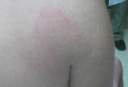
Erythema migrans in Lyme disease.[40] [Figure caption and citation for the preceding image starts]: Erythema migransCourtesy of Christian Speil, Southern Illinois University School of Medicine, Springfield, IL [Citation ends].
Dengue fever presents with diffuse skin flushing of the face, neck, and chest, evolving into a maculopapular or rubelliform rash that can affect the whole body. Flushing may blanch when affected skin is pressed. Haemorrhagic signs include petechiae, purpura, or a positive tourniquet test (performed by inflating a blood pressure cuff to a point midway between systolic and diastolic blood pressures for 5 minutes; the test is positive if ≥10 petechiae per square inch appear on the forearm).
Petechial rash with bacterial infections, infectious endocarditis, Rocky Mountain spotted fever, leukaemia, and some viral infections.
Malar (butterfly) rash in SLE.
Erythema marginatum in rheumatic fever.
Erythema nodosum with sarcoidosis and tuberculosis.
Cutaneous lesion at site of inoculation with cat-scratch disease.
Diaphoresis with medication-related reactions.
Severe pruritus in lymphomas and uraemia.
Scarlatiniform rash in scarlet fever; sandpaper-like generalised erythematous rash with discrete skin-coloured papules; linear petechial streaks in skinfolds, particularly axillary, inguinal, and antecubital fossae (Pastia lines).
Eyes
Conjunctivitis with Kawasaki disease and viral infections.
Papilloedema in patients with a cerebral abscess, meningitis, encephalitis.
Retinal haemorrhages are observed in child abuse that may be associated with factitious disorder imposed on another.
Uveitis in Crohn's disease, ulcerative colitis, and leptospirosis.
Proptosis may be present in patients with thyroid storm.
Inability to make tears suggests paediatric autonomic disease.
Severe retro-orbital pain on eye movement or with little pressure applied to the eyeball is common in dengue fever.
Joints
Swollen joints suggest septic arthritis, JIA, SLE, rheumatic fever, leukaemia, lymphoma, serum sickness, endocarditis, inflammatory bowel diseases.
Other musculoskeletal signs
Osler nodes (painful tips of fingers) with bacterial endocarditis, a sign of septic emboli.
Subcutaneous nodules on extensor aspects of extremities in rheumatic fever.
Thyroid
Goitre with bruit suggests thyrotoxicosis.
Lymphadenopathy
Isolated cervical lymphadenopathy can be seen in patients with Kawasaki disease and cat-scratch disease.
Disseminated lymphadenopathy is observed with infectious mononucleosis, CMV, toxoplasmosis, leukaemia, lymphomas, tick-borne illnesses, and some inflammatory/vasculitic illnesses.
Most bacterial infections give rise to regional lymphadenopathy.
The ulceroglandular form of tularaemia typically presents with lymphadenopathy.
Respiratory examination
Crackles (rales) are observed with pneumonia, viral infections, and tuberculosis.
Pleuritis may be present in inflammatory/vasculitic disorders and malignancies.
Cardiovascular examination
A heart murmur may be present with bacterial endocarditis.
Pericardial friction rub may be detected in patients with pericarditis associated with several inflammatory/vasculitic disorders.
Abdominal examination
Superficial tenderness can be found with cystitis, pyelonephritis, hepatic abscess (right upper quadrant), typhoid fever, and inflammatory bowel disease.
Hepatomegaly and/or splenomegaly suggests typhoid fever, infectious mononucleosis, CMV, toxoplasmosis, endocarditis, myocarditis, hepatic abscess, malaria, tuberculosis, JIA, or SLE.
An abdominal mass may be palpated due to lymphadenopathy.
Perianal fissures and abscesses are found in Crohn's disease.
Testicular pain may be present in patients with leukaemia, metastases from other malignancies, or septic emboli following endocarditis or in brucellosis.
ENT examination
Sinus tenderness may be found in sinusitis.
Bulging tympanic membrane may indicate otitis media.
Erythema of the tonsils may be present in patients with acute tonsillitis. Purulent exudate on the tonsillar surface may be present, particularly when it is caused by group A beta-haemolytic streptococci.
Cervical lymphadenopathy, pharyngeal erythema with exudates, palatal petechiae, and a red, swollen (strawberry) tongue suggest scarlet fever.[41]
Nervous system
Kernig's and Brudzinski's signs can be found in meningitis.
Bulging fontanelle.[11]
Focal neurological deficits can present with any cerebral space-occupying lesion such as an abscess, malignancy, or septic emboli from heart vegetation (endocarditis).
Diagnostic testing
The need for diagnostic testing in the child depends on the baseline risk of an underlying infection or other serious aetiology.[42] Because viruses or bacteria cause the overwhelming majority of fevers in children who present acutely, initial evaluation is often focused on detecting a source of infection. A meticulous physical examination may often reveal an obvious source, and the identification of a focal infection may obviate the need for additional testing.
With the exception of neonates and young infants, in whom the physical examination is less reliable, the approach can be more selective if the child has a non-toxic appearance after initial examination.[43][44] More rigorous testing is required in patients who are at higher risk of infection and in whom history and examination findings are limited. Traditionally, this has led to different approaches to diagnostic testing based on age; immunisation status also plays a role in pretest risk stratification.
Coronavirus disease 2019 (COVID-19)
Over 90% of children are asymptomatic or have a mild coryzal illness. However, moderate to severe illness has been reported in children.[27] It is important to note that children may have signs of pneumonia on chest imaging despite having minimal or no symptoms.[28] Co-infection with other respiratory viruses is common.
Physical examination may detect fever (with or without chills/rigors) and obvious cough and/or difficulty breathing in cases of severe illness. Auscultation of the chest may reveal inspiratory crackles, rales, and/or bronchial breathing in patients with pneumonia or respiratory distress. Patients with respiratory distress may have tachycardia, tachypnoea, or cyanosis accompanying hypoxia.
Molecular testing is required to confirm the diagnosis. Diagnostic tests should be performed according to guidance issued by local health authorities and should adhere to appropriate biosafety practices.
Chest x-ray or CT chest should be considered. Ground-glass opacities on CT are present in 60% of patients. Consolidation with surrounding halo signs is considered a typical feature of paediatric infection.[28]
Evaluation of fever without source (acute presentation)
The vast majority of children who present acutely with 'fever without source' (or 'fever of unclear source') have underlying infections, typically requiring urgent evaluation and empirical treatment (especially in young children).
Management of sepsis in children first requires prompt recognition, so it is imperative to consider the possibility of sepsis whenever a child presents with a fever. Criteria to identify sepsis and septic shock in children and young people under the age of 18 years have been developed.[45] Treatment guidelines have been produced by the Surviving Sepsis Campaign and remain the most widely accepted standards.[15]
Children up to 2 months of age
Children in this age group are at higher risk of bacterial infections than older children. History and physical examination are poorly sensitive in young children; those with serious bacterial illness may have normal physical examinations.
Initial laboratory tests[46][47][48]
FBC (including a peripheral WBC count) is often ordered in the evaluation of febrile neonates and young infants, but the discriminatory value is insufficient to differentiate between patients with serious bacterial infection and non-bacterial infection. While an abnormally high or low peripheral WBC count increases the concern for bacteraemia or meningitis, it is an imperfect screening tool, and the decision to obtain blood culture and spinal fluid should not depend on the results.[49][50] Erythrocyte sedimentation rate (ESR) and CRP are also used as screening tools for infection, but have the same limitations as WBC count. Elevated procalcitonin levels may be a better predictor of serious bacterial infection in these patients.[51][52]
Blood culture.
Urine for rapid testing (obtained by either urethral catheterisation or suprapubic aspiration) and urine culture.
Cerebrospinal fluid (CSF) studies including a CSF culture.
Chest x-ray: usually indicated only in the presence of respiratory symptoms.[53]
Stool analyses: indicated only in the presence of diarrhoea.[11]
Children over 2 months of age
History and physical examination are more informative in older patients and the diagnostic approach can therefore be more selective, depending on the history and symptomatology.
The following tests may be considered:
FBC (including a peripheral WBC count): although commonly ordered in the evaluation of a febrile child, the test is not sensitive or specific enough to help make a definitive diagnosis. As with younger children, ESR, CRP, and procalcitonin are sometimes used as screening tools for infection.
Urine microscopy and culture: urinary tract infections (UTIs) are common in girls and uncircumcised boys up to 24 months of age. Positive dipstick test result for any of urine leukocyte esterase, nitrites, leukocyte count can be used to make a preliminary diagnosis of urinary tract infection in febrile patients.[54] Enhanced urinalysis and culture is requested; a urine culture should be obtained when starting antibiotics.[54] A combination of findings (including history of UTI, temperature ≥39.0°C (102°F), fever duration of more than 24 hours, absence of another source of fever, suprapubic tenderness, uncircumcised male infant) increases the probability of UTI.[54][55]
Blood culture: serious infections can be occult, especially in younger children. Febrile children up to 24 months of age with temperatures >39.0°C (102°F ) and <2 pneumococcal vaccinations are at higher risk for bacteraemia.[36]
CSF testing: should be reserved for children with history or physical examination findings suggestive of CNS infection.
Stool studies: indicated in patients suspected of having inflammatory bowel disease and typhoid fever.
Chest x-ray: pneumonia presenting as fever without any respiratory symptoms is unusual.[56][57] A chest x-ray should be obtained if examination findings are suggestive of hypoxaemia or significant respiratory distress (e.g., tachypnoea, dyspnoea, retractions, grunting, nasal flaring, apnoea, or altered mental status), if there is high fever, fever longer than 48 hours, or tachycardia or tachypnoea disproportionate to fever, and for those patients failing initial antibiotic therapy.[53][54][58]
Evaluation of fever of unknown origin (subacute presentation)
Fever of unknown origin is not well defined in children and has been historically used to describe a subacute presentation of a single illness of at least 3 weeks duration during which a fever >38.3°C (100.9°F) is present for most days and the diagnosis is unclear after 1 week of intense investigation.[1]
The most common causes are infections, inflammatory/vasculitic disorders, and malignancies. These children require a more deliberate, comprehensive, and prolonged evaluation, and frequently do not need urgent empirical therapy. There is an increased emphasis on unusual presentations of common illness and on esoteric causes.
Investigations to be considered include:
FBC and peripheral smear: the WBC count is a non-specific test but if elevated, suggests infection, while a low WBC count can be seen in viral illnesses or sepsis. Anaemia is present in certain conditions associated with fever (e.g., infective endocarditis, malaria). Elevated platelet count is non-specific and may be seen acutely in a variety of illnesses; similarly, thrombocytopenia can be seen with severe bacterial or viral illnesses. Peripheral smear may demonstrate abnormal cells suggestive of a haematological malignancy or may detect malarial parasites.
ESR, CRP, and procalcitonin: non-specific parameters of infection and/or inflammation.[52][59]
Blood culture.
Urinalysis and urine culture.
Electrolytes, blood urea nitrogen, creatinine, liver function tests.
Serological tests (Epstein-Barr virus, CMV, HIV).
Chest x-ray.
Nucleic acid amplification tests (to identify M tuberculosis).
Additional tests (as indicated)
Stool testing
Bone marrow examination
Lumbar puncture
Serum antinuclear antibody
Imaging studies
Abdominal imaging
Sinus imaging
Mastoid imaging
Labelled WBC scanning
Echocardiography.
Specific diagnostic criteria
Fever ≥5 days without any other explanation plus 4 principal criteria with coronary artery abnormalities.
Principal criteria
Changes in extremities (acute erythema of palms or soles, or oedema of hands and feet; periungual peeling of digits seen in weeks 2 and 3 of illness)
Polymorphous exanthema
Bilateral bulbar conjunctival injection without exudates
Changes in lips and oral cavity (erythema, cracked lips, strawberry tongue, diffuse injection of oral and pharyngeal mucosa)
Cervical lymphadenopathy (>1.5 cm diameter) usually unilateral.
These symptoms may have resolved by the time of presentation. It is important to enquire whether the child has had these symptoms since the onset of fever, even if they are not present at the time of assessment.[11]
Rheumatic fever (Jones criteria)[61]
If preceded by recent group A streptococcal infection, diagnosis can be made by meeting 2 major, or 1 major and 2 minor, criteria. A presumptive diagnosis can be made if chorea or carditis is the only manifestation. Furthermore, because recurrence is difficult to diagnose in patients with previous rheumatic fever or rheumatic heart disease, these patients need to meet either 1 major or 2 minor criteria in the setting of recent group A streptococcal infection.
Major criteria: carditis, polyarthritis, subcutaneous nodules, chorea, erythema marginatum.
Minor criteria: fever, arthralgia, elevated acute-phase reactants (ESR, CRP), prolonged PR interval on ECG, elevated or rising anti-streptolysin O titre or DNase, previous rheumatic fever.
Streptococcal toxic shock syndrome[62]
A confirmed case meets the clinical criteria plus isolation of group A Streptococcus from a normally sterile site.
Criteria include hypotension (<5th percentile for age for children <16 years of age) and 2 or more of the following:
Renal impairment
Coagulopathy
Liver involvement
Acute respiratory distress syndrome
Generalised rash that may desquamate
Soft-tissue necrosis including necrotising fasciitis, myositis, or gangrene.
Staphylococcal toxic shock syndrome[62]
A confirmed case requires presence of fever, hypotension, diffuse erythroderma, desquamation (unless the patient dies before desquamation can occur), and involvement of at least 3 organ systems.
A probable case is in a patient who is missing one of the characteristics of the confirmed case definition.
Use of this content is subject to our disclaimer