Approach
The diagnosis of croup depends upon a careful history and physical examination. The key features are the characteristic sudden-onset, seal-like barky cough, often accompanied by stridor and chest wall (intercostal) or sternal indrawing. Symptoms are typically worse at night and increase with agitation.
There may be a history of prior non-specific upper respiratory tract symptoms (coryza, non-barky cough, mild fever), although the seal-like barky cough may also present abruptly with no preceding illness. Although not essential to the diagnosis, there is commonly a hoarse voice.
Clinical presentation
Presentations may range from mild symptoms to impending respiratory failure.[19] The physician should look out for the following symptoms and signs according to severity:
Mild: seal-like barky cough but no stridor or sternal/intercostal recession at rest
Moderate: seal-like barky cough with stridor and sternal recession at rest; no agitation or lethargy
Severe: seal-like barky cough with stridor and sternal/intercostal recession, associated with agitation or lethargy
Impending respiratory failure: increasing upper airway obstruction, sternal/intercostal recession, asynchronous chest wall and abdominal movement, fatigue, and signs of hypoxia (pallor or cyanosis) and hypercapnia (decreased level of consciousness secondary to rising PaCO₂). The degree of chest wall recession may diminish with the onset of respiratory failure as the child tires.
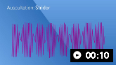 Stridor
Auscultation sounds: Stridor
The clinician must consider the differential diagnosis during the physical examination. In particular, if epiglottitis is suspected, examination of the oropharynx or manipulation of the neck is contraindicated as it may precipitate further airway obstruction.
Work-up
Croup is largely a clinical diagnosis.[20] X-ray of the anteroposterior and lateral neck is not performed in a child presenting with typical symptoms and signs of croup. The steeple sign (narrowed trachea) is a classic finding on anteroposterior view, but is not always present. Radiological studies are contraindicated if there is clinical suspicion of epiglottitis or bacterial tracheitis, as manipulation of the neck region and agitation may precipitate further airway obstruction. If the clinical picture is atypical for these conditions, soft-tissue radiographs of the neck may provide helpful information to support an alternative diagnosis. Any x-ray should be performed with considerable care and personnel equipped to support the airway in the event of worsening obstruction.
Use of this content is subject to our disclaimer