Approach
Traditionally, vitamin B3 deficiency has been diagnosed when clinical features compatible with pellagra are evident.[80] Specialised biochemical assessment of niacin nutritional status is cumbersome, and lacks sensitivity and specificity. Nicotinic acid concentration in the urine and serum is usually low and does not change appreciably with changes in nicotinic acid intake. The first clinical signs of vitamin B3 deficiency occur shortly after the urinary metabolites of niacin fall to very low levels.[80]
Pellagra may be difficult to identify as its main symptoms of dermatitis, diarrhoea, and dementia occur in an unpredictable order. Diagnosis may also be complicated by the fact that vitamin B3 deficiency can coexist with other B vitamin deficiencies, including vitamin B2 and B6, which present with similar symptoms.[4] Furthermore, pellagra has become rare in developed countries and may thus be overlooked, and its diagnosis is difficult in the absence of dermatitis.[81]
Once a diagnosis has been confirmed, the underlying cause of the deficiency must be determined. It is necessary to assess the risk of primary or secondary vitamin B3 deficiency. A dietary history should be taken to help determine whether it is an isolated primary dietary deficiency or part of a multinutrient deficiency state.
Clinical features
The signs and symptoms of pellagra can easily be misinterpreted.[82] The key features are characterised by diverse dermatological, neurological, and gastrointestinal (GI) clinical manifestations that do not develop in any specific order.[8]
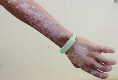
Cutaneous features begin with a symmetrical, bilateral, and well-defined erythematous rash on exposed areas of skin sensitive to sunlight. The appearance is similar to sunburn.[81] Dermatitis can rapidly become severe, characterised by hyperpigmentation, bullous lesions, and desquamation on exposed parts of the body (e.g., face, neck, and arms).[46] Casal's necklace occurs in advanced presentations and is characterised by erythema, hyperpigmentation, and scales around the neck.[28] Dermatitis is not always present in pellagra.[7][8][56] Other dermal symptoms include pruritic eruptions on the dorsum of the hand and distal forearm; erythema and slight scaling of the face, neck, and upper limbs; and nasolabial seborrhoea and vulvovaginitis. Repeated exposure to ultraviolet (UV) light leads to butterfly erythema on the face, persistent dermatitic plaques, telangiectasia, and poikilodermatous changes.[83]
A range of neuropsychological features are seen in some patients; including anxiety, depression, intermittent stupor, insomnia, vertigo, memory loss, paranoia, hallucinations, delirium, altered gait, peripheral neuropathy, weakness and fatigue.[81] While folate, vitamin B12, and vitamin B6 are known to be important for brain health, there is a general lack of controlled trial research into the effects of the remaining B vitamins (thiamine, riboflavin, biotin, pantothenic acid, niacin) on brain function.[84]
Pellagrous encephalopathy includes symptoms of confusion, oppositional hypertonus, and oppositional myoclonus.[7][32][56][85] Cogwheel rigidity, primitive reflexes (grasping and sucking), ataxia, hallucinations, and incontinence have also been reported.
GI features include cachexia, diarrhoea or constipation, nausea and vomiting, anorexia and weight loss, and heartburn. Inflammation of the oral cavity progresses to the oesophagus, and eventually to the entire GI tract.
Tongue and oral features include glossitis, cheilosis, stomatitis, gingivitis, and fissures on the sides of the tongue.[8][57] It should be noted that these features are difficult to differentiate from those of folic acid, thiamine, B2, B6, and B12 deficiencies.[1]
Other clinical features include achlorhydria and angular palpebritis.[Figure caption and citation for the preceding image starts]: Typical dermatitis of pellagra on sun-exposed skin (extensor aspect of forearm and dorsum of hand) that is clearly demarcated from unexposed skinFrom the collection of Demetre Labadarios [Citation ends].
 [Figure caption and citation for the preceding image starts]: Typical dermatitis of pellagra on sun-exposed skin (face and anterior neck) that is clearly demarcated from unexposed skinFrom the collection of Demetre Labadarios [Citation ends].
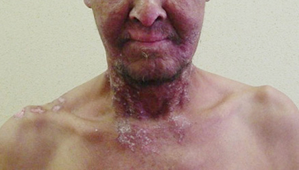 [Figure caption and citation for the preceding image starts]: Typical dermatitis of pellagra on sun-exposed skin (posterior neck and ear helices) that is clearly demarcated from unexposed skinFrom the collection of Demetre Labadarios [Citation ends].
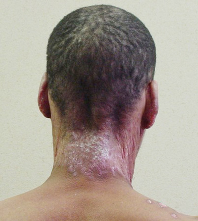
Risk factors
Vitamin B3 deficiency should be suspected in certain groups of patients.
Dietary deficiency and malnutrition may result from the consumption of a corn-based, low-protein diet, or a vegan diet (no animal products) with few niacin and/or tryptophan sources. It may also occur in very poor, homeless, or displaced people; in those with an eating disorder (e.g., anorexia nervosa, bulimia nervosa); in those with chronic alcohol use disorder; in those who have a genetic defects that affects nutrition; and those who are under stress.[21]
Malabsorption may be secondary to Crohn's disease, Roux-en-Y bariatric surgery, or other causes of the short bowel syndrome.
Some medications, such as isoniazid and rifampicin for the treatment of tuberculosis, pyrazinamide, ethionamide, fluorouracil, mercaptopurine, hydantoins, phenobarbital, chloramphenicol, oestrogen-containing oral contraceptives, and penicillamine, may also lead to vitamin B3 deficiency.
Other conditions associated with vitamin B3 deficiency include Hartnup's disease, carcinoid syndrome, and HIV infection.
Confirmation of vitamin B3 deficiency
Serum tryptophan levels are below the normal reference range.[86] This is associated with the consumption of a diet low in tryptophan.
Urinary metabolites of niacin are very low.[80] Adults normally excrete 20% to 30% of niacin intake as N-methylnicotinamide and 40% to 60% as N-methyl-2-pyridone-5-carboxamide in the urine. The urinary ratio of these metabolites is an indicator of niacin status. A ratio of <1 (mg/g creatinine) is indicative of niacin deficiency.[80]
Histopathological findings on skin biopsy may include non-specific dermatitic changes, perivascular lymphohistological infiltrates in the upper dermis, mild oedema in the papillary dermis, capillary dilatation, and dermal fibrosis. Epidermal changes include early subcorneal blisters; pallor of the upper epidermis; hyperkeratosis; parakeratosis; and epidermal atrophy, detachment, and necrosis.[87][88][89]
Photosensitivity testing involves the exposure of healthy skin to minimal erythema doses of UV light. Photosensitivity reactions include erythema, swelling, and blistering on exposed parts of the body (i.e., face, upper back, chest, extensor aspect of forearm), as well as pruritic, burning, or tingling sensations.
Characteristic ECG changes may occasionally be seen: diffuse ST-T changes, ST depression and diphasic or flattened T waves, and inversions in leads II, III, aVF, and V1-V5. Niacin therapy normalises the ST-T changes.
Diagnosis can be confirmed with a therapeutic trial of niacin 3 times daily for 5 days. In the presence of vitamin B3 deficiency, improvement in cutaneous symptoms usually occurs within 48 hours of treatment.[8]
Investigation of underlying cause
If primary dietary deficiency is deemed unlikely, other aetiologies leading to secondary vitamin B3 deficiency, based on the past medical and drug histories, should be specifically investigated.
A FBC may reveal microcytic (iron deficiency) or macrocytic (vitamin B12 deficiency) anaemia in malnutrition. The MCV will also be elevated in chronic alcohol use disorder. LFTs may be deranged and the gamma-GT elevated in chronic alcohol use disorder. Serum albumin and protein levels may be low in malnutrition.
Measures of iron (serum total iron, haemoglobin, iron-binding capacity, ferritin), thiamine (transketolase activity, thiamine pyrophosphate), riboflavin (riboflavin coefficient), pyridoxine (serum pyridoxal-5-phosphate), cyanocobalamin (serum cyanocobalamin), and folic acid (serum folic acid, red cell folic acid) deficiency may reveal concomitant nutritional deficiency.
Examination of the stool may reveal malabsorption. Hartnup's disease is indicated by a high neutral aminoaciduria level, and carcinoid syndrome is indicated by an elevated serum serotonin and urinary 5-hydroxyindoleacetic acid. HIV antibodies are positive in HIV infection. There is some evidence for the involvement of niacin in the development of neurodegenerative diseases such as Alzheimer's disease and Parkinson's disease, but this evidence is inconclusive.[74][75]
Use of this content is subject to our disclaimer