Approach
The history and physical examination are key components for the diagnosis of varicose veins. This is complemented by mandatory duplex ultrasonography to assess for reflux, not only in the varicosities but also in the truncal veins.
History
Patients may present with varied symptoms, and many may be asymptomatic. Some of the more common symptoms include heaviness or fatigue in the lower extremities and ankle oedema, especially with prolonged standing. Burning and itching of the skin over the veins, restless legs, and leg cramps (usually nocturnal) may also be present. Symptoms are generally worse during pregnancy or menstruation. Patients will often find relief with elevation of the leg or while wearing compression hosiery. During the evaluation, patients should be questioned regarding previous interventions on their veins as well as history of trauma, deep vein thrombosis, ulcerations, or bleeding from varicosities. Most varicose veins do not progress to complications such as ulceration.[13]
Examination
With the patient standing, the skin is examined visually and by palpation for irregularities and bulges consistent with varicose veins. The extent, size, and location of the dilated veins should be noted, along with skin changes such as haemosiderin depositions, lipodermatosclerosis (C4b), and areas of active (C6) or healed (C5) ulceration. Superficial venous thrombosis presents as severe pain, erythema (and superficial pigmentation), and vein induration. Hyperpigmentation, lipodermatosclerosis (area of induration because of fibrosis of subcutaneous fat), or ulcers are indicative of long-standing venous insufficiency. [Figure caption and citation for the preceding image starts]: Varicose veinsFrom the collection of Maureen K. Sheehan, MD; used with permission [Citation ends].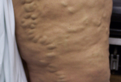 [Figure caption and citation for the preceding image starts]: Haemosiderin depositionFrom the collection of Maureen K. Sheehan, MD; used with permission [Citation ends].
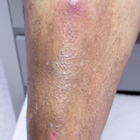
Imaging
Duplex ultrasound imaging uses B mode imaging and Doppler assessment to diagnose venous insufficiency by evaluating valvular function in various segments of the truncal veins as well as in the varicosities.[14] B mode imaging is used to rule out deep vein thrombosis and persistent obstruction in the venous system. Venous duplex ultrasound scans are performed in the standing position and take approximately 20 minutes per leg. Repeated calf compressions are used to identify reflux of blood.
Use of this content is subject to our disclaimer