Approach
Diagnosis is clinical. Wood lamp examination is helpful in determining histological subtype of melasma. Skin biopsy is done only if the clinical examination suggests another diagnosis, such as post-inflammatory hyperpigmentation or hyperpigmentation due to drugs or to exogenous ochronosis.
History
Clues supporting a diagnosis of melasma include female sex, darker skin types, history of recent pregnancy or of oral contraceptive pill use, and sun exposure. Dyschromia that follows inflammation or use of perfumes or cosmetics is less likely to be melasma and more likely to be Riehl melanosis or post-inflammatory hyperpigmentation.[3]
Physical examination
Muddy brown to grey hyperpigmentation in a typical distribution (i.e., centrofacial, malar, mandibular) suggests a diagnosis of melasma. It can sometimes affect different areas such as the forearms and neck. [Figure caption and citation for the preceding image starts]: MelasmaFrom the collection of Dr Laura Ferris [Citation ends].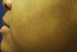
Wood lamp examination
If a diagnosis of melasma is suspected, Wood lamp examination can be helpful in determining whether it is primarily epidermal (accentuated by Wood lamp examination) or dermal (not accentuated by Wood lamp examination). Mixed dermal/epidermal melasma will show spotty accentuation with Wood lamp examination. This test can be helpful in choosing a treatment modality, as epidermal melasma is more likely to respond to topical therapy, whereas dermal melasma is more likely to require laser treatments. However, some studies suggest that classification by Wood lamp may not correlate with biopsy results.[13]
Reflectance confocal microscopy (RCM)
A tool that may be considered for the classification of melasma in the future. It is a non-invasive test for skin imaging and microscopic analysis. Using melanin and melanosomes as contrast for RCM images, the technique is able to identify histological changes associated with melasma. It allows for semi-quantification of the amount of pigment in each layer of the skin and can be used to classify the histological type of melasma without the need for an invasive procedure such as a skin biopsy.[19] One case report reported on the successful use of RCM to assess melasma in 20 female patients at baseline and 3-month follow-up after treatment.[20]
Use of this content is subject to our disclaimer