Approach
Characteristic history and examination findings are often sufficient to diagnose the condition.
History
AK presents typically in a man with light-coloured skin, >40 years old, having spent a lot of time outdoors since childhood without sun protection.
AKs are more prevalent in people living at lower latitudes, and in people with genetic DNA instability and melanin deficiency (e.g., autosomal recessive inherited type 1 and type 2 albinism, and xeroderma pigmentosum).[15][16][17][18]
Physical examination
Single or multiple lesions in sun-exposed areas including forehead, bald areas of the scalp, ears, lower lip, and dorsum of the hands and forearms. The lesions are commonly skin-coloured, yellowish, or erythematous, ill-defined, irregularly shaped, small (1 to 5 mm), rough, scaly macules or plaques.[50]
There may be mild pruritus, irritation, or bleeding if the lesions have been scratched.
Other presentations include:
Scaly lesions with a hyperkeratotic surface >1 mm in height (hyperkeratotic AKs)[50]
Well-defined, scaly, brown lesions resembling solar lentigo (pigmented AKs)[1]
Lesions resembling seborrhoeic keratosis, melanocytic naevus, and early malignant melanoma (spreading pigmented AKs)[5]
Skin-coloured, papillomatous, elevated wart-like papules (verrucous AKs)
Plaques with very mild scale over very thin shiny skin (atrophic AKs)
Violaceous well-defined papules with fine white lines on the surface (lichen planus-like or lichenoid AKs)
Hypertrophic conical-shaped protuberances growing from the surface of the skin (cutaneous horn)[50]
Scaly red roughness with induration, fissuring, and ulceration of the lower lip to the commissures (actinic cheilitis).[1][4]
Other skin signs of sun damage may also be present.[Figure caption and citation for the preceding image starts]: Regular actinic keratosisFrom the collection of the Department of Dermatology and Cutaneous Surgery, University of Miami Miller School of Medicine [Citation ends]. [Figure caption and citation for the preceding image starts]: Actinic cheilitisFrom the collection of the Department of Dermatology and Cutaneous Surgery, University of Miami Miller School of Medicine [Citation ends].
 [Figure caption and citation for the preceding image starts]: Hyperkeratotic actinic keratosisFrom the collection of the Department of Dermatology and Cutaneous Surgery, University of Miami Miller School of Medicine [Citation ends].
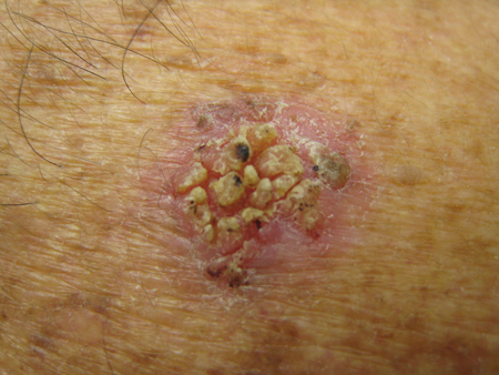
Investigations
Dermoscopy is performed only if the clinical examination findings are not typical of AK.
AK lesions have a strawberry pattern on dermoscopy.[51] This consists of an erythematous background, pink-to-red pseudo-network, thin undulated vessels surrounding hair follicles, white-to-yellow scale, and hair follicles filled with yellowish keratotic plugs, surrounded by a white halo. Possible early signs of progression of facial AKs to squamous cell carcinoma (SCC) include dotted vessels surrounding hair follicles, starburst pattern, and coalescence of keratotic follicles.[52]
Skin biopsy can be performed if the clinical examination and dermoscopy findings are not typical of AK and there is a suspicion of progression to SCC; however, biopsy is rarely required. Major clinical criteria associated with the progression of AKs to SCC are induration/inflammation, diameter >1 cm, rapid enlargement, bleeding, erythema, and ulceration. Minor criteria associated with the progression of AKs to SCC are pigmentation, palpability, pain, pruritus, and hyperkeratosis.[53]
Use of this content is subject to our disclaimer