Approach
Hyperkalaemia can be difficult to detect clinically because associated symptoms are typically vague. Hyperkalaemia is for the most part detected as an incidental laboratory finding.
The history is most useful in identifying conditions, such as renal failure or adrenal insufficiency, commonly associated with hyperkalaemia, or determining whether medications, such as potassium-sparing diuretics or potassium supplements, are in use. An obvious cause of hyperkalaemia can usually be detected based on the history, once transcellular shifts of potassium, sampling conditions, and decreased renal excretion have been taken into account.
Clinical features
Low-level hyperkalaemia, in the range of 5.0 to 6.0 mmol/L (5.0 to 6.0 mEq/L), is almost always asymptomatic. Values of serum potassium >7.0 mmol/L (>7.0 mEq/L) are more often symptomatic by way of muscle weakness and are evident on ECG. Muscle weakness is uncommon below serum potassium values of 7.0 mmol/L (7.0 mEq/L). Electrocardiographic changes with hyperkalaemia can progress rapidly from asymptomatic findings to life-threatening arrhythmias.
Features that should be elicited from the history include common causes of hyperkalaemia, including: acute or chronic renal failure with or without a high potassium intake; muscle trauma; chemotherapy for a rapidly proliferating tumour; and poorly controlled diabetes with significant hyperglycaemia.[23][24] Patients with recent significant muscle injury, prolonged seizures, and/or a history of having exercised excessively in a warm environment should be suspected of having some degree of rhabdomyolysis. A symptom complex characterised by weight loss, fatigue, excessive skin pigmentation, cold intolerance, hypotension, and a tendency to develop hyponatraemia and hypoglycaemia should arouse suspicion of Addison's disease. A complete drug history should also be obtained. Drug history should include intake of high doses of potassium supplements and the use of any of several different medications, such as: non-steroidal anti-inflammatory drugs, ACE inhibitors, angiotensin-receptor blockers, heparin, pentamidine, ciclosporin (cyclosporine), tacrolimus, trimethoprim, and/or potassium-sparing diuretics (e.g., spironolactone, eplerenone, canrenone, triamterene, and amiloride).[4][5][7][8][10][11][13][15]
Step-by-step consideration of potential causes
It is not unusual for there to be multiple contributing factors causing hyperkalaemia.
In a patient with normal renal function, a normal ECG and/or a history of a haematological disorder, the physician should take steps to exclude pseudohyperkalaemia.[39]
Pseudohyperkalaemia is identified by a serum (clotted blood) potassium that is >0.4 mmol/L (>0.4 mEq/L) higher than the potassium level in a simultaneously-obtained plasma (nonclotted blood) sample. Spurious hyperkalaemia can be caused by difficult venepuncture, prolonged transit time and poor storage conditions.[28] A quick review of a recent FBC, seeking out significant leukocytosis (>100,000 x 10⁹/L) or thrombocytosis (>500,000 x 10⁹/L), will establish whether these causes of pseudohyperkalaemia are present.
Once pseudohyperkalaemia has been ruled out, the pathophysiological mechanism of cellular redistribution is considered and is a likely cause if there is significant hyperglycaemia, and/or a medication history of mannitol, arginine hydrochloride (rarely used to treat significant metabolic alkalosis), suxamethonium (succinylcholine), digoxin, and/or beta-blocker.
If both pseudohyperkalaemia and cellular redistribution have been ruled out as causes, the next consideration is an imbalance between potassium intake and excretion. Consideration of potassium intake relative to the level of renal function becomes most important. Dietary potassium intake can be difficult to establish based on recall; only when salt substitutes or potassium supplements are being used can a high intake be definitively established. Reduced renal function alone seldom results in hyperkalaemia without there being a significant dietary intake. In those instances when serum potassium values are much higher than would be expected for the level of renal function, the differentials of hyporeninaemic hypoaldosteronism or hypoaldosteronism should be investigated. The following tests should be done for confirmation: plasma renin activity, plasma cortisol, and plasma aldosterone.
Interpretation of serum potassium concentrations
There is a limited correlation between the serum potassium concentrations and total body potassium stores.
Spurious laboratory values, termed pseudohyperkalaemia, can falsely increase the serum potassium value; accordingly, it is important to consider repeating the test for confirmation. Pending confirmatory testing, the serum potassium value in question should be viewed as being correct and appropriate treatment measures instituted.
Investigations
All patients presenting with hyperkalaemia should have the following initial tests: basic metabolic panel (including serum potassium, glucose, bicarbonate, urea, and serum creatinine), serum calcium, FBC, and an ECG.
Subsequent tests performed depend on the clinical findings, and include:
urine dipstick creatine kinase, if rhabdomyolysis is a possibility
cortisol and aldosterone levels, if Addison's disease is suspected
arterial blood gases, if metabolic acidosis needs to be established
serum digoxin level, if the patient is receiving digoxin or suicide is known to have been attempted with digoxin ingestion
urine pH, to evaluate for inappropriately elevated value (pH >5.5) in the setting of metabolic acidosis, suggesting renal tubular acidosis
transtubular potassium gradient, to evaluate for various forms of impaired distal tubular secretion of potassium (determination of the transtubular gradient for potassium requires urine and plasma samples for potassium and osmolality)
plasma renin activity, usually elevated in pseudohypoaldosteronism
17-hydroxyprogesterone, if 21-hydroxylase deficiency is suspected in newborns.
Electrocardiogram
An ECG should be performed in all patients with significant hyperkalaemia. ECG findings are usually progressive and include first degree heart block (prolonged PR interval >0.2 seconds), flattened or absent P waves, peaked (tented) T waves (i.e. T wave larger than R wave in >1 lead), ST-segment depression, widened QRS (>0.12 seconds), ventricular tachycardia, bradycardia and eventually cardiac arrest (pulseless electrical activity, ventricular fibrillation/paroxysmal ventricular tachycardia, asystole).[28][Figure caption and citation for the preceding image starts]: ECG changes in patients with hyperkalaemiaBMJ 2009; 339:b4114. Copyright ©2009 by the BMJ Publishing Group [Citation ends].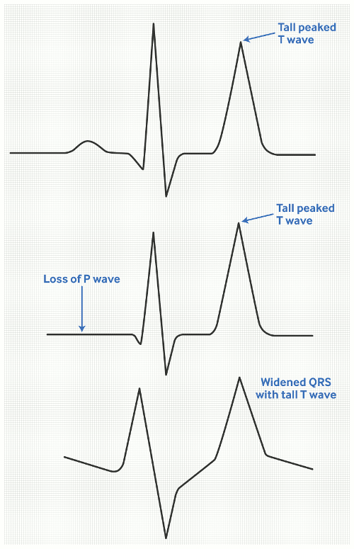
Hyperkalaemia with decreased urine potassium excretion
A 24-hour urine potassium excretion of <20 mmol/L (<20 mEq/L) will distinguish renal from extra-renal causes (potassium excretion >40 mmol/L [>40 mEq/L]) of hyperkalaemia. However, urinary potassium measurements may prove difficult to interpret, since multiple factors influence these values independent of level of renal function.
For values that are difficult to interpret, the transtubular potassium gradient is of some use. The transtubular potassium gradient (TTKG = [K+] urine/(U/P)osm/plasma K+) corrects urinary potassium for changes in osmolality that occur with absorption of water in the collecting duct. A value consistently <7 suggests impaired distal tubular secretion of potassium (secondary either to hypoaldosteronism or to aldosterone resistance); a value >10 favours increased intake and intact distal tubular handling of potassium.[40]
Hyperkalaemic periodic paralysis
Hyperkalaemic periodic paralysis should be considered when there is intermittent muscle weakness in the setting of cold exposure, ethanol use, potassium administration, and/or a high-potassium diet. With weekly determinations of potassium values, high normal values can occasionally be detected during attack-free intervals, which then allows for a presumptive diagnosis.
Use of this content is subject to our disclaimer