Images and videos
Images


Thoracic outlet syndrome
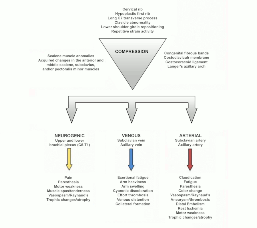
Schematic diagram summarising the factors involved in thoracic outlet compression and the resulting signs and symptoms
From the collection of Dr Robert W Thompson (adapted from Dr Chaney Stewman); used with permission
See this image in context in the following section/s:

Thoracic outlet syndrome
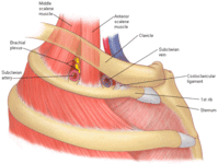
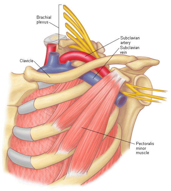
Lateral view of the neurovascular structures traversing the thoracic outlet, with the clavicle above and the first rib below. The costoclavicular space is the second major site of neurovascular compression
Reprinted with permission from Elsevier
See this image in context in the following section/s:
Thoracic outlet syndrome
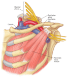
The neurovascular structures pass behind the pectoralis minor muscle, a third major area of neurovascular compression. The pectoralis minor muscle is a shoulder protractor, which can overcome the rhomboids. The shoulder retracts and alters the thoracic outlet, contributing to muscle imbalance and compression of the brachial plexus
Reprinted with permission from Elsevier
See this image in context in the following section/s:

Thoracic outlet syndrome
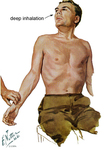
The Adson maneuver tightens the anterior and middle scalene muscles, decreasing interspace and magnifying pre-existing compression of the brachial plexus and subclavian artery. The patient takes and holds a deep breath, extends the neck fully, and turns the head toward the affected side. Obliteration or decrease in the radial pulse indicates neurovascular compression at the thoracic outlet
Reprinted with permission from Netter Images
See this image in context in the following section/s:
Thoracic outlet syndrome
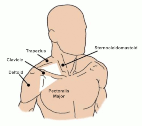
Surface landmarks of the thoracic outlet
From the collection of Dr. Robert W. Thompson; used with permission
See this image in context in the following section/s:
Thoracic outlet syndrome
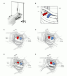
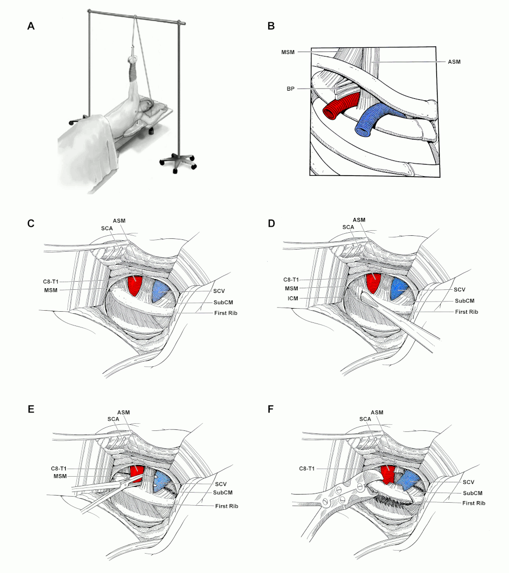
Transaxillary first rib resection and scalenectomy. (A): Patient position. (B): Anatomical features of the thoracic outlet as viewed from the lateral approach. (C): Exposure of the first rib high in the axilla, with the associated muscles and subclavian vessels visualised. (D): Dissection along the periosteum of the first rib. (E): Middle scalene and subclavius muscles are divided and the anterior scalene is encircled prior to its division. (F): Division of the posterior first rib. ASM: anterior scalene muscle; BP: brachial plexus; C8-T1: C8 and T1 cervical nerve roots; MSM: middle scalene muscle; SCA: subclavian artery; SCV: subclavian vein; SubCM: subclavius muscle
Arnaoutakis GJ, et al. Atlas of Vascular Surgery and Endovascular Therapy: Anatomy and Technique. Chaikof EL, Cambria RP, editors. Chapter 16. Transaxillary rib resection for thoracic outlet syndrome. Elsevier Saunders: Philadelphia; 2014. Pages 193-203; used with permission.
See this image in context in the following section/s:

Thoracic outlet syndrome
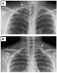
Cervical ribs. Chest radiographs demonstrating a patient with a partial cervical rib on the right side (A) and a patient with a complete cervical rib on the left side (B)
From the collection of Robert W. Thompson, MD; used with permission
See this image in context in the following section/s:

Thoracic outlet syndrome
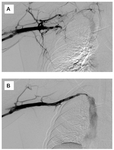
Upper extremity venography. (A): Initial venogram showing thrombotic occlusion of the axillary-subclavian vein. (B): Completion venogram after catheter-directed thrombolysis and suction thrombectomy, with restoration of a patent axillary-subclavian vein and residual venous stenosis at the level of the first rib
From the collection of Dr. Robert W. Thompson; used with permission
See this image in context in the following section/s:

Thoracic outlet syndrome
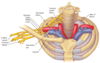
The subclavian vein and artery pass over the first rib and under the clavicle. The brachial plexus traverses the top of the bony circle to join the artery. The apex of the pleura (cupula) is shown on the left side
Reprinted with permission from Elsevier
See this image in context in the following section/s:
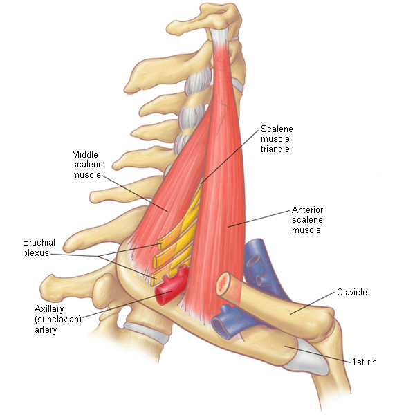
Thoracic outlet syndrome
The scalene muscle triangle is the first major level of neurovascular compression
Reprinted with permission from Elsevier
See this image in context in the following section/s:

Thoracic outlet syndrome
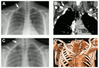
Subclavian artery aneurysms. (A and B): Plain chest radiograph showing complete cervical rib on the right side (arrow) and an image from CT angiogram showing a subclavian artery aneurysm arising immediately past the point of arterial compression by the cervical rib (arrow). (C and D): Plain chest radiograph showing complete cervical rib on the right side (arrow) and a 3-D reconstruction from a CT angiogram demonstrating a subclavian artery aneurysm immediately past the point of arterial compression by the cervical rib (oval highlight). CR: cervical rib
From the collection of Dr. Robert W. Thompson; used with permission
See this image in context in the following section/s:
Use of this content is subject to our disclaimer