Differentials
Medial collateral ligament (MCL) sprain
SIGNS / SYMPTOMS
The pop sound at injury is less common and true dynamic instability is rare, unless it is a complete tear or with associated injuries. May be able to continue activity if minor sprain. No significant haemarthrosis in isolated MCL sprain, although reactive effusion is not uncommon. Tender over MCL course/insertion. MCL stress testing reveals laxity and/or pain. Additionally, Lachman's and pivot shift tests are negative. Spontaneous healing and full return of function is the rule.[59]
INVESTIGATIONS
MRI reveals fluid around or injury to MCL; ACL appears intact. Subsequent x-rays may reveal calcification along previously injured MCL (Pellegrini-Stieda disease).
Posterior collateral ligament (PCL) sprain
SIGNS / SYMPTOMS
Often able to continue activity but knee does not feel right subjectively to the patient. Instability may be more subtle, with knee effusion typically smaller and less noticeable by the patient. Lachman's, pivot shift, anterior drawer tests are negative, but the posterior drawer test is positive. External rotation recurvatum test will show posterolateral corner instability. Unlike with ACL tears, non-operative approach often allows return to full activity.[60][Figure caption and citation for the preceding image starts]: Lachman's manoeuvreFrom the personal collection of Philip H. Cohen [Citation ends]. [Figure caption and citation for the preceding image starts]: Anterior and posterior drawer test starting positionFrom the personal collection of Philip H. Cohen [Citation ends].
 [Figure caption and citation for the preceding image starts]: External rotation recurvatum test.From the personal collection of Philip H. Cohen [Citation ends].
INVESTIGATIONS
MRI reveals disrupted PCL, and an intact ACL.
Lateral collateral ligament (LCL) sprain
SIGNS / SYMPTOMS
Local swelling is common, but significant effusion is rare. There is tenderness over the lateral collateral ligament and/or bony insertion. A complete tear often results in a palpable gap. Spontaneous healing and full return of function is the rule.[61]
INVESTIGATIONS
MRI reveals fluid around or disruption to LCL, with intact ACL.
Meniscal tear
SIGNS / SYMPTOMS
Meniscal injury may also occur with ACL tear or other significant injury. Main symptom is pain at affected joint line and stiffness. Effusion typically develops over 1 to 2 days, but rapid haemarthrosis may occur if large tear in vascular portion of meniscus.[62][63]
Physical examination manoeuvres have poor accuracy, but joint line tenderness is common, as is pain with rotational manoeuvres (e.g., positive McMurray's test). [Figure caption and citation for the preceding image starts]: McMurray's flexion varusFrom the personal collection of Philip H. Cohen [Citation ends].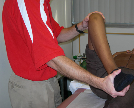 [Figure caption and citation for the preceding image starts]: McMurray flexion in valgusFrom the personal collection of Philip H. Cohen [Citation ends].
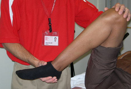 [Figure caption and citation for the preceding image starts]: McMurray's external varusFrom the personal collection of Philip H. Cohen [Citation ends].
 [Figure caption and citation for the preceding image starts]: McMurray's external valgusFrom the personal collection of Philip H. Cohen [Citation ends].
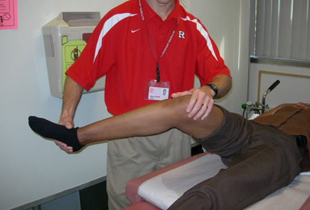
INVESTIGATIONS
MRI has excellent sensitivity for meniscal tears, but intra-substance degenerative changes may be misread as acute tears.[64]
Posterior capsular sprain
SIGNS / SYMPTOMS
Small effusion and stiffness common, along with painful extension. ACL and other ligaments are stable.[65]
INVESTIGATIONS
Imaging rarely necessary. MRI may show effusion, bone bruising, abnormal signal at posterior capsule, but no frank ligament disruption.
Patellar subluxation or dislocation
SIGNS / SYMPTOMS
Patient feels severe pain around patella; may see kneecap may appear displaced to the side. Haemarthrosis occurs, with difficulty in weight bearing, and a sensation of the kneecap being unstable. Examination usually reveals haemarthrosis, tenderness at patella and medial retinaculum, and apprehension when examiner attempts to displace patella laterally. ACL testing should be normal.[66]
INVESTIGATIONS
X-rays may reveal dislocation if not yet reduced, or patellar fracture. Lateral subluxation and abnormal patellofemoral relationship often noted. MRI often reveals bone bruising, and may show chondral injury, osteochondral fracture, or loose body.[67]
Use of this content is subject to our disclaimer