Images and videos
Images

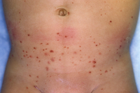
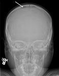

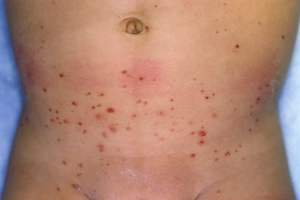
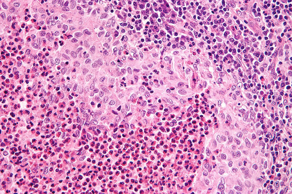
Langerhans cell histiocytosis
Skull x-ray showing lytic bone lesion in the right posterior parietal area of the skull
From the personal collection of Oussama Abla, MD
See this image in context in the following section/s:
Langerhans cell histiocytosis
Langerhans cell histiocytosis rash on an infant's abdomen
Reproduced with permission from Science Photo Library
See this image in context in the following section/s:
Langerhans cell histiocytosis
Very high magnification micrograph of Langerhans cell histiocytosis. H&E stain. It is characterised by Langerhans-type histiocytes that have a reniform (kidney-shaped) nucleus and stain with S100 and CD1a
Nephron. Reproduced under a creative commons license CC BY-SA 3.0: https://creativecommons.org/licenses/by-sa/3.0/deed.en
See this image in context in the following section/s:
Use of this content is subject to our disclaimer