Approach
Cervical spondylosis may be clinically evident either from the patient's presenting symptoms (i.e., neck pain, cervical radiculopathy, or cervical myelopathy) or from an incidental finding during an evaluation of other symptoms.[7][16]
For patients with cervical neck pain there are several symptoms that should be considered red flags and that indicate the need for further investigation to exclude important differential diagnoses:[16][27]
History of recent fall or trauma to the head or neck
Unexplained weight loss
Severe, intractable pain or severe local tenderness
Cervical lymphadenopathy
Unexplained fever, especially in diabetic patients
History of cancer
History of chronic steroid use.
Presenting symptom of primarily axial neck pain
Axial neck pain includes components of direct pain (particularly experienced in the posterior aspects of the neck) and referred or indirect components.[2][6][7] Referred components include paraspinal muscle spasm initiated from joint motion, with secondary pain arising from sustained muscle contraction and stiffness. Other referred components include occipital pain and tension headaches.
Axial neck pain can exist in any axial neck muscle, including scalenes (anterior scalene syndrome), trapezius and interscapular muscles, and paraspinal muscles extending from the occiput to the lumbar region, where axial muscle spasm can spread.[20]
The degree of neck pain with cervical spondylosis is more subjective than noted on examination, with mild to moderate paraspinal muscle spasm, nearly preserved range of motion, and tenderness in the paraspinal muscle groups (scalenes, trapezius, interscapular muscles, occipital, etc.).[2][6][7]
For a single episode only, a therapeutic trial of treatment may be sufficient to establish the diagnosis. Further diagnostic tests may not be warranted.
Cervical radiographs are indicated for patients with severe neck pain, chronic neck pain, or pain with a history of trauma or neck surgery (recent or previous).[5] With a history of malignancy or neck surgery, guidelines suggest an MRI is more appropriate.[5]
Presenting symptom of radiating arm pain, with or without axial neck pain
The diagnosis of cervical spondylotic radiculopathy depends on the presence of radiating arm pain (distal to the shoulder); the pattern following ≥1 well-defined dermatomes; mild weakness in the muscles innervated by a specific nerve root; and reflex changes for a single level if appropriate.[7][28] There is rarely any significant loss of function because the nerve roots overlap considerably.
[Figure caption and citation for the preceding image starts]: Chart showing average dermatome size and location. Radicular pain is usually confined to a single dermatomeFrom the 20th US edition of Gray's Anatomy of the Human Body; used with permission [Citation ends].
Depending on the correlation with physical and neurological examination and the duration (particularly >4 to 6 weeks), further diagnostic tests may be helpful, though not initially critical unless the history and neurological examination do not converge.[24]
In most cases, cervical radiographs are less helpful than proceeding directly to a magnetic resonance imaging (MRI) scan if the symptoms persist. If an MRI is not possible (e.g., implanted metal), then a computed tomography (CT) cervical spine scan is the next most appropriate study.[5] If a cervical CT scan with no contrast suggests spinal cord abnormalities and an MRI is not possible, the next step is a CT cervical spine scan with intrathecal contrast (CT myelography) to obtain more detail about spinal cord and nerve changes.[5]
If a single nerve root is difficult to distinguish (particularly considering the similar patterns between C6 and C7), then occasionally a nerve test, such as electromyography (EMG) or nerve conduction velocity (NCV), can be helpful. The EMG specifically tests muscles for signs of denervation that can arise between the spinal cord and muscle, while the NCV helps to localise specific areas of nerve compression or damage.
However, for localisation of a single nerve root, a specific nerve block (laterally at the nerve exit from the spine) is more definitive as a diagnostic and sometimes therapeutic intervention.[29] The EMG/NCV can help discern other entities with weakness and/or sensory loss that may mimic a radiculopathy, particularly focal mononeuropathy (i.e., diabetes) and peripheral nerve compression syndromes.
Presenting symptoms of loss of upper extremity function, with or without axial neck pain
Degenerative cervical myelopathy (DCM) remains a poorly defined syndrome.[10][27][30][31] It includes often painless reduction of neurological function arising from compression of the spinal cord due to a variety of degenerative changes in the cervical spine.[13] These changes can include bony overgrowth at any of the disc or facet joints (i.e., osteophytes), ligamentous changes or calcification, instability, or change in alignment of the joints (i.e., kyphotic deformity).[1][3][4] The extent of radiological involvement may not correlate with the extent of neurological involvement.
From the history of weakness and/or numb, clumsy hand syndrome, a correlative neurological examination is important to identify whether this is a subjective or objective finding.[13] The most common initial signs of myelopathy are mild hand weakness (particularly in the intrinsic muscles of the hand, such as the interossei), loss of coordination in hand function (such as typing), and mild gait ataxia.[27] More severe symptoms can include profound weakness of the hands, bowel and bladder difficulties, and severe gait ataxia.[13]
There is rarely a significant loss of proximal muscle strength in the arms or legs, and such loss would be a clear red flag beyond DCM. Cervical myelopathy can worsen acutely with injury to the cervical spine, although there is usually a pre-injury history of loss of function.
If there are neurological signs suggesting loss of function beyond a single nerve root, a cervical MRI is important to distinguish the specific nature of any neural compression. Cervical MRI scans clearly show spinal cord compression and deformation with usually internal T2 (white) signal changes in the spinal cord.[3][Figure caption and citation for the preceding image starts]: A single level of spinal cord compression with T2 changes, on cervical sagittal T2 sequence in the presence of symptomatic degenerative cervical myelopathyDennis A. Turner, MA, MD [Citation ends].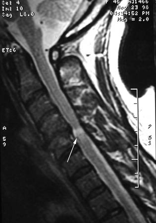 [Figure caption and citation for the preceding image starts]: Previous spinal cord compression at C3/4 on sagittal T2 MRI, with residual T2 changes, and new compression at C2/3 and C6/7, with T2 changesDennis A. Turner, MA, MD [Citation ends].
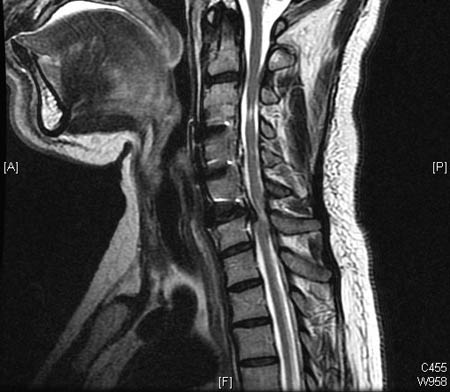
A cervical MRI with and without contrast may be considered, which will indicate alternate causes, including malignancy, infection, inflammatory diseases, and intrinsic myelopathy or internal damage to the spinal cord (i.e., multiple sclerosis, amyotrophic lateral sclerosis).[5] If the weakness and/or sensory loss is unilateral, then another consideration is brachial plexopathy (neuralgic amyotrophy) due to spontaneous viral neuronitis, often with initial neck pain.
Because the neurological abnormalities arise from spinal cord compression and upper motor neuron dysfunction rather than nerve compression, EMG or NCV is a negative diagnostic test in cervical myelopathy, as nerve studies primarily demonstrate lower motor neuron changes. However, the EMG/NCV can be helpful to differentiate cervical myelopathy from more peripheral disorders, such as ulnar neuropathy or carpal tunnel syndrome, which can mimic many of the symptoms.
Asymptomatic but noted to have cervical spondylosis on diagnostic evaluation for some other condition
This is a common form of presentation. The scan itself is interpreted as abnormal and the patient seeks to know the significance of the scan findings.[1][4] The patient's presenting symptom commonly resolves by the time of consultation regarding the scan findings.
A correlation between the patient's history and neurological and physical examination is critical to decide whether further diagnostic tests are warranted.[2][6] If clear neurological abnormalities are noted, then an MRI scan is suggested if possible.[32] If the patient has had an MRI scan, a thorough discussion with the patient may be critical to decide whether to suggest treatment in an asymptomatic patient, particularly when specific risks may arise from this treatment.
Common asymptomatic findings include vertebral body haemangiomas (which are congenital, although can rarely expand over time), disc or facet degenerative changes (which are age related in severity and extent), disc bulging, annular tear, and presence of a visible central spinal canal (often mislabelled as a syrinx).[1][4]
Use of this content is subject to our disclaimer