Approach
Mechanism of injury
Patient description of the mechanism of injury is an essential part of the history. For example, most anterior cruciate ligament tears are non-contact twisting injuries. Some of the main aspects of the mechanism of injury to be investigated include location of contact, location of pain, history of previous injury, and areas of anaesthesia/dysaesthesia.[6][7]
Location of contact
Blow to the anterior aspect of knee
Blow to hyperextended knee: may cause anterior cruciate ligament (ACL) or posterolateral corner injuries.
Blow to flexed knee (such as in a dashboard injury): may cause posterior cruciate ligament (PCL) injury.
Blow to the lateral aspect of knee (valgus injury)
This injury would stretch the medial-sided knee structures, risking injury to the superficial medial collateral ligament.
Blow to the medial aspect of knee (varus injury)
This would increase the stress on the outside of the knee, and may cause a posterolateral corner injury.
Non-contact twisting injuries
Patellar subluxation or dislocation may occur when the knee is twisted in extension. This is especially true in patients with patella alta, where the patella is abnormally high in relation to the femur and is not well engaged within the trochlear groove.
If the knee is twisted while in flexion, meniscus or ACL tears are important considerations.
Location of pain
Medial knee pain
For acute knee injuries: consider a medial meniscus injury, articular cartilage injury of the medial compartment, or medial collateral ligament (MCL) injury.
For chronic injuries: consider medial compartment arthritis, pes anserine bursitis, and a degenerative medial meniscal tear.
Lateral knee pain
For acute knee injuries: differentials include a lateral meniscus tear, lateral compartment articular cartilage injury, or posterolateral corner injury (to include the fibular collateral ligament).
For chronic knee injuries: consider lateral compartment arthritis, biceps femoris bursitis, iliotibial band friction syndrome and a degenerative lateral meniscus tear.
Anterior knee pain
For acute injuries: patellar subluxation or dislocation and patellar or trochlear groove chondral injuries are possible diagnoses. Patellar tendon rupture should be considered in patients 30 to 45 years old; quadriceps tendon rupture is a possibility in patients 45 to 60 years old.
For chronic injuries: consider patellofemoral joint chondromalacia (arthritis) and patellar tendonitis.[6]
Posterior knee pain
For acute knee injuries: differentials including posterior capsule injuries, posterior cruciate ligament injury, or posterior horn meniscal tears.
For chronic knee injuries: posterior horn meniscal tears or a Baker's (popliteal) cyst are considered.
History of previous injuries
Many patients have symptoms secondary to either a previous injury or previous surgery. It is important to recognise if any changes in the swelling or motion, when compared with the normal contralateral knee, are acute or chronic. Patients who have had previous ligament injuries, with or without surgery, are at an increased risk for re-injuring structures and causing other secondary injuries. In addition, patients who have had previous resections of their menisci for tears are at a high risk of developing arthritis of the same compartment, which could lead to pain and swelling with activities.
Areas of anaesthesia/dysaesthesia or strength deficiency
It is important to assess for any new areas of sensory change or weakness around the patient's knee and lower extremity after a knee injury.
Direct blows to the anterior aspect of the knee can cause sensory changes in the infrapatellar branch of the saphenous nerve, resulting in altered sensation on the anterolateral aspect of the knee.
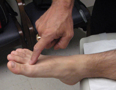
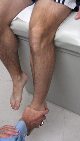
Blows to the anteromedial knee or a varus or hyperextension injury can cause a stretch injury to the common peroneal nerve. These patients will have decreased sensation in the first dorsal web space and lateral aspect of their foot, compared with the contralateral leg. In addition, they may have motor weakness of the extensor hallucis longus, small toe extensors, and foot evertors, and ankle dorsiflexion. [Figure caption and citation for the preceding image starts]: First dorsal web space sensation testing (common peroneal nerve)From the personal library of Dr LaPrade; used with permission [Citation ends].
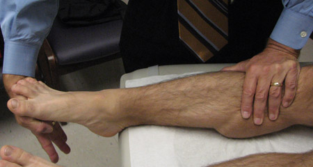
 [Figure caption and citation for the preceding image starts]: Lateral foot sensation testing (superficial peroneal nerve)From the personal library of Dr LaPrade; used with permission [Citation ends].
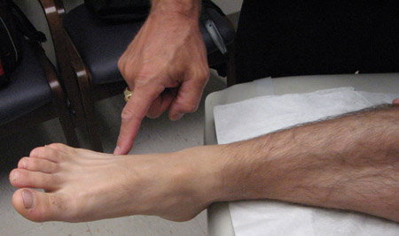 [Figure caption and citation for the preceding image starts]: Extensor hallucis longus strength testingFrom the personal library of Dr LaPrade; used with permission [Citation ends].
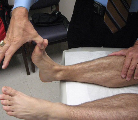 [Figure caption and citation for the preceding image starts]: Small toe extensors strength testingFrom the personal library of Dr LaPrade; used with permission [Citation ends].
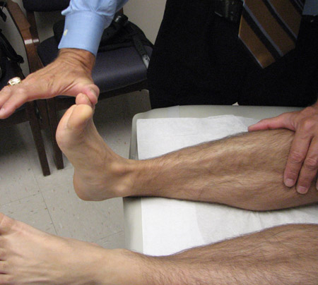 [Figure caption and citation for the preceding image starts]: Foot eversion strength testingFrom the personal library of Dr LaPrade; used with permission [Citation ends].
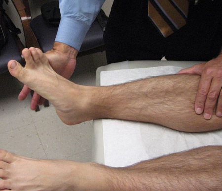 [Figure caption and citation for the preceding image starts]: Dorsiflexion strength testingFrom the personal library of Dr LaPrade; used with permission [Citation ends].
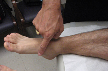
Patients with injuries to the medial aspect of their knee, including lacerations, may have decreased sensation along the medial aspect of the leg and foot consistent with an injury to the saphenous nerve (i.e., medial foot). [Figure caption and citation for the preceding image starts]: Medial foot sensation (saphenous nerve)From the personal library of Dr LaPrade; used with permission [Citation ends].
In patients with knee dislocation or a multi-ligament injury, it is necessary to assess the function of the tibial nerve by assessing the sensation on the sole of the foot, and ankle plantar flexion and inversion strength. [Figure caption and citation for the preceding image starts]: Plantar flexion strength testingFrom the personal library of Dr LaPrade; used with permission [Citation ends].
 [Figure caption and citation for the preceding image starts]: Foot inversion strength testingFrom the personal library of Dr LaPrade; used with permission [Citation ends].
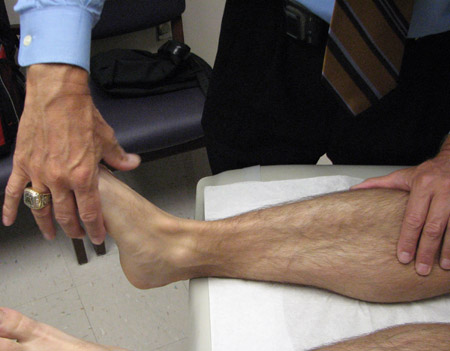
Assessment of quadriceps strength and full extension is recommended as an indication of patellar tendon injuries, patellofemoral dysfunction, and injuries involving the femoral nerve.
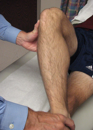
Assess hamstring strength as an indication of injuries to the tibial nerve and acute hamstring tears. [Figure caption and citation for the preceding image starts]: Quadriceps strength testingFrom the personal library of Dr LaPrade; used with permission [Citation ends].
 [Figure caption and citation for the preceding image starts]: Hamstring strength testingFrom the personal library of Dr LaPrade; used with permission [Citation ends].
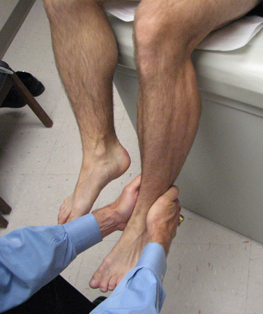
General physical examination
A thorough physical exam is necessary to document specific pathologies associated with acute or chronic knee injuries. In the majority of cases, it is possible to determine the pathology that is present. Following a series of examination steps on a regular basis is important to obtain a thorough and complete physical exam.[6]
Acute effusion
For young patients with an acute traumatic effusion, the most common injury is an anterior cruciate ligament tear, followed by meniscal tears and patellar subluxations/dislocations.[8]
In older patients, the pathology may involve meniscal tears, tibial plateau fractures, aggravations of underlying arthritic processes, or ligament injuries.
Joint warmth or redness may be indicative of an infection or an inflammatory process.
Chronic knee effusions
In chronic effusions (including those with obvious trauma), causes include an underlying osteoarthritic process, inflammatory process, infection, or tumour.
Subtle signs of effusions include having the patient flex the knee and looking for some anterior protrusion of the margins of the fat pad on either side of the patellar tendon, or in palpating the knee posteriorly to look for signs of a Baker's cyst. A Baker's cyst usually indicates that there is swelling elsewhere within the joint and that this fluid has leaked out posteriorly in the knee in the space between the direct arm of the semimembranosus tendon and the medial head of the gastrocnemius tendon.
Deformities of the knee
Deformities of the knee can be due to patellar dislocation (almost always lateral), fractures, knee dislocations, or penetrating wounds.
Knee dislocations are classified according to the tibial location on the femur.
Other deformities can be due to patellar tendon ruptures, with proximal migration of the patella and a visible step-off deformity distal to the patella.
Quadriceps tendon ruptures show distal migration of the patella, with an obvious deformity proximal to the patella.
Loss of skin integrity
Lacerations around the knee are particularly concerning. They may indicate a penetrating wound into the knee joint, an open fracture, or an open knee dislocation.
Lacerations that extend into the prepatellar bursa are very concerning due to the high risk of infection
Complete or incomplete range of motion
Compare the range of motion of the injured knee to the normal contralateral knee.
Causes of lack of full extension include a bucket handle tear of the meniscus or trauma to the anterior aspect of the knee (retropatellar fat pad contusion, anterior horn meniscal tear, or fracture).
A lack of full flexion can be due to an injury to the posterior horn of the menisci, a knee effusion limiting full flexion, or other posterior knee pathologies.[6]
Document the location of the patients' pain when they are having their range of motion assessed. Anterior knee pain with flexion of the knee usually indicates a patellofemoral source of discomfort. Posterior knee pain with flexion of the knee has a higher likelihood of being due to meniscal pathology. Patients who have a lack of full extension in chronic injuries usually have some underlying arthritis, or may have post-injury or post-surgical changes.[Figure caption and citation for the preceding image starts]: Hip external rotationFrom the personal library of Dr LaPrade; used with permission [Citation ends].
 [Figure caption and citation for the preceding image starts]: Hip internal rotationFrom the personal library of Dr LaPrade; used with permission [Citation ends].
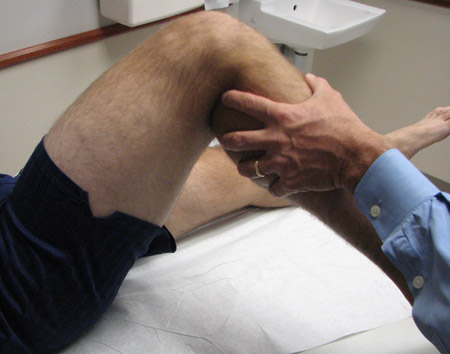 [Figure caption and citation for the preceding image starts]: Log rolling testFrom the personal library of Dr LaPrade; used with permission [Citation ends].
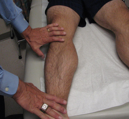 [Figure caption and citation for the preceding image starts]: Deep flexion of the kneeFrom the personal library of Dr LaPrade; used with permission [Citation ends].
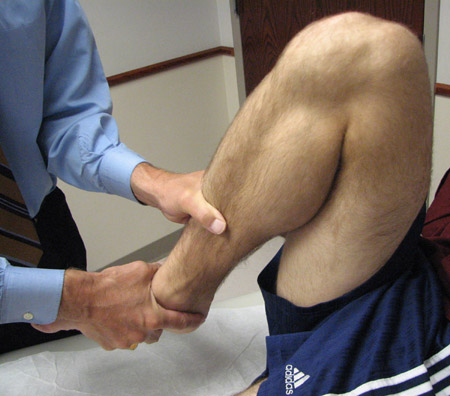
Assess the range of motion of the hip joint.
External rotation recurvatum test
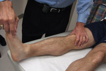
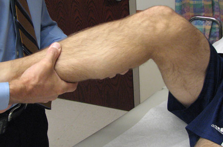
The great toe is lifted up while some gentle downward pressure is held on the anterior thigh (to hold it against the examination table) to see if there is any increase in recurvatum (i.e., hyperextension) compared with the contralateral side, usually measured in centimeters of heel height.
An increase in recurvatum is almost always due to a severe knee injury, which can include a combined ACL and posterolateral corner injury, a possible severe posteromedial knee injury with an MCL injury, or a knee dislocation.[9][10][Figure caption and citation for the preceding image starts]: External rotation recurvatum testFrom the personal library of Dr LaPrade; used with permission [Citation ends].
Physical examination for patellofemoral joint injuries
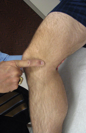
The medial suprapatellar plica is palpated by rolling the medial plica over the medial femoral condyle with the index finger to assess for any associated pain.[11] This can commonly be a source of knee pain with patellofemoral dysfunction.[Figure caption and citation for the preceding image starts]: Palpation of medial plicaFrom the personal library of Dr LaPrade; used with permission [Citation ends].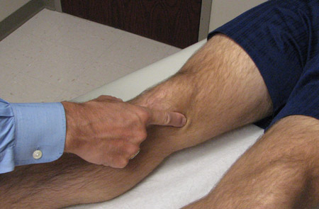 [Figure caption and citation for the preceding image starts]: Palpation of inferior pole of patellaFrom the personal library of Dr LaPrade; used with permission [Citation ends].
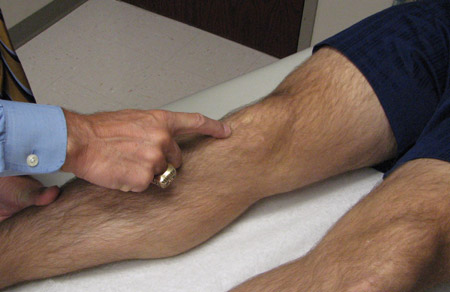 [Figure caption and citation for the preceding image starts]: Palpation of pes anserine bursaeFrom the personal library of Dr LaPrade; used with permission [Citation ends].
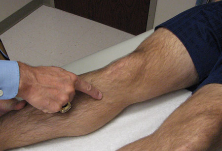 [Figure caption and citation for the preceding image starts]: Palpation of prepatellar bursaeFrom the personal library of Dr LaPrade; used with permission [Citation ends].
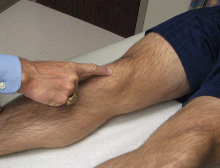 [Figure caption and citation for the preceding image starts]: Palpation of superior pole of patellaFrom the personal library of Dr LaPrade; used with permission [Citation ends].
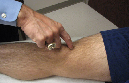 [Figure caption and citation for the preceding image starts]: Palpation of semimembranosus bursaFrom the personal library of Dr LaPrade; used with permission [Citation ends].
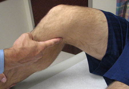
Patellar translation/instability assessment
The lateral patellar apprehension test is usually performed at 45° of knee flexion. In this test, any increase in lateral translation that causes pain to the patient and a reactive contraction of the quadriceps muscle, when compared with the contralateral side, is indicative of a previous patellar dislocation.[11]
Increased medial subluxation of the patella is almost always due to an excessive lateral release. In this circumstance, the patella can subluxate or dislocate medially in the initial flexion arc of the knee before the patella becomes engaged in the trochlear groove. This instability is usually in the first 0° to 40° of knee flexion.[11][Figure caption and citation for the preceding image starts]: Lateral patellar translationFrom the personal library of Dr LaPrade; used with permission [Citation ends].
 [Figure caption and citation for the preceding image starts]: Medial patellar translationFrom the personal library of Dr LaPrade; used with permission [Citation ends].
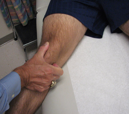
Assessment of retropatellar crepitation with translation of the patella in the trochlear groove
The patient needs to be totally relaxed. The patella is translated medially and laterally in the trochlear groove, with the knee flexed to approximately 20° to 45°, to determine if there is any associated retropatellar crepitation and any associated pain with this test. In addition, the superior and inferior poles of the patella are rolled over the trochlear groove to assess for crepitation at these portions of the patella, which may indicate some localised chondromalacia. It is important to assess for pain or discomfort during this manoeuvre.[Figure caption and citation for the preceding image starts]: Rolling superior and inferior poles of patella within trochlear grooveFrom the personal library of Dr LaPrade; used with permission [Citation ends].
 [Figure caption and citation for the preceding image starts]: Rolling superior and inferior poles of patella within trochlear grooveFrom the personal library of Dr LaPrade; used with permission [Citation ends].
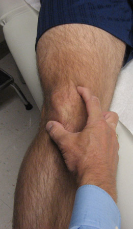
Palpation of retropatellar fat pad
The retropatellar fat pad can be a source of pain either post-injury or due to scar tissue following surgery. Palpation of the retropatellar fat pad is performed along the edges of the patellar tendon, and can help to determine if this location is a source of pathology. [Figure caption and citation for the preceding image starts]: Palpation of lateral patellar fat padFrom the personal library of Dr LaPrade; used with permission [Citation ends].
 [Figure caption and citation for the preceding image starts]: Palpation of medial patellar fat padFrom the personal library of Dr LaPrade; used with permission [Citation ends].
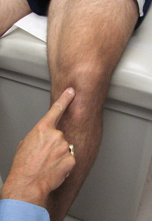
Physical examination for anterior cruciate ligament injuries
Lachman's test
Can be used to differentiate an ACL tear from other knee injuries. The test is performed between 20° and 30° of knee flexion. One hand stabilises the thigh, while the other hand is placed just distal to the tibial tubercle. An anterior translation force is then applied to the tibia to see if there is any increase in anterior motion. Increases in anterior translation are most commonly due to a torn anterior cruciate ligament.[12]
Note that there may be a false negative Lachman's test in patients that do not totally relax or in patients who have sustained a bucket handle tear of either of the menisci.
A pseudo-Lachman's test can be obtained when there is uncertainty where the starting point of the tibia is on the femur. In some patients with posterior cruciate ligament injuries, the knee may be subluxed posteriorly and there may be a false sense of increased anterior translation, when in fact the starting point is more posterior in the knee. To a lesser extent, a pseudo-Lachman's test can be obtained in the face of a posterolateral corner injury when the knee starts off in a more posteriorly translated position. To differentiate a pseudo-Lachman's from a true positive Lachman's test, there is usually a hard endpoint felt at the end of the Lachman's test rather than a soft endpoint. Further, the endpoint of the tibia in relation to the femur is not as anterior as one would expect when there is an ACL tear.[Figure caption and citation for the preceding image starts]: Lachman's testFrom the personal library of Dr LaPrade; used with permission [Citation ends].
Anterior drawer test
Performed with the knee between 80° and 90° of knee flexion and the foot in neutral rotation.[6] In this test, an anterior translation force is applied to see if there is any increase in the anterior translation of the tibia on the femur, compared with the normal contralateral knee.
A positive anterior drawer test can be indicative of an ACL tear or an ACL tear combined with a posterior capsular knee injury. In addition, patients who have had the posterior horn of their menisci excised or injured may have a positive anterior drawer test.
The anterior drawer test by itself is not as pathognomonic for an ACL tear as the Lachman's test.
Anterior drawer test in external rotation
Performed with the knee flexed to between 80° and 90°. The foot is then externally rotated approximately 15°. In this test, the knee is evaluated to determine if there is increased anterior medial rotation of the tibia on the femur. A positive anteromedial drawer test is indicative of a combined MCL and posteromedial joint injury. It is important to differentiate this test from the posterolateral drawer test, which looks at posterolateral rotation of the tibia on the femur.
Pivot shift test
Assesses the amount of anterolateral rotatory instability of the knee with an anterior cruciate ligament tear. In effect, this test is looking for the amount of anterolateral rotation of the tibia that occurs in the knee near full extension.[13]
The knee is positioned close to full extension. A valgus force is then applied to the knee, along with an internal rotation force. It is very important to make sure that the patient is completely relaxed. One way to help patients relax is to apply a slight traction force (distally) to the leg to slightly open the knee joint. The examiner then observes for anterolateral subluxation of the tibia on the femur. The knee is then flexed while applying the same forces, and one observes for a reduction of the anterolateral subluxation of the tibia on the femur. This is usually observed with an appreciable 'clunk' noise heard by the examiner. A positive pivot shift test is usually indicative of an anterior cruciate ligament tear.[Figure caption and citation for the preceding image starts]: Pivot shift testFrom the personal library of Dr LaPrade; used with permission [Citation ends].
Pitfalls in pivot shift test assessment
False negative pivot shift tests are very common; examiner experience, as well as guarding by the patient, may contribute. Thus, it is important to make sure that the patient is completely relaxed while performing this test.[13] The examiner should avoid gripping the leg tightly; a more gently applied force may sometimes help the patient to relax sufficiently to determine if there is an anterior cruciate ligament tear.
In patients who have a bucket handle tear of the meniscus, there may be a false negative pivot shift test because the tibia is not able to sublux anteriorly on the lateral aspect of the femur. This pitfall is especially true for bucket handle tears of the lateral meniscus.
Physical examination for medial collateral ligament injury and associated valgus laxity
Valgus laxity is a true rotation laxity; it is not pure medial joint line opening.
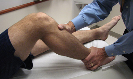
Assessment for valgus laxity
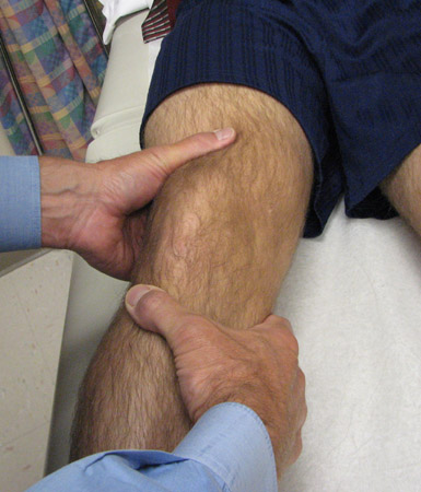
The patient should be relaxed and positioned with the thigh on the examining table and the leg over the side of the bed. One hand on the foot applies the valgus force, to assess for the combined medial opening and rotational opening. The other hand has the fingers directly over the joint line, to assess for medial joint line opening.[14] It is important to differentiate true medial joint line opening from increased motion due to joint line collapse and pseudolaxity, which can be found in patients with medial compartment arthritis.
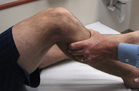
Increased valgus opening in extension is usually indicative of a severe medial-sided joint injury combined with an injury to one of the cruciate ligaments. Increased valgus opening at 30° can be indicative of a partial or complete MCL tear, depending upon whether there is a stable endpoint present after one applies the valgus force.[14][Figure caption and citation for the preceding image starts]: Valgus stress testFrom the personal library of Dr LaPrade; used with permission [Citation ends].
Assessing medial collateral ligament injuries
It is important to establish which portion of the superficial medial collateral ligament and deep MCL may have been injured. The portion of the superficial medial collateral ligament that attaches to the femur is called the meniscofemoral portion. This portion, more commonly injured, has a better prognosis in the case of isolated injuries.
The meniscotibial portion, which attaches to the tibia usually heals, but has a lesser chance of healing compared with the meniscofemoral portion.
It is useful for both the physiotherapist and the athlete to recognise the location of the MCL injury, as this may be useful in determining the type of physiotherapy and/or bracing to include in the post-treatment regimen.
Pitfalls in assessing for valgus instability
In patients with open growth plates, a growth plate (physeal) fracture may mimic increased varus or valgus joint line opening. Therefore, it is important to utilise stress x-rays in this population.[6]
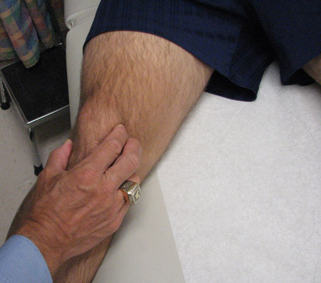
In older patients with seemingly increased amounts of joint line gapping, there may be associated fractures around the knee, including tibial plateau fractures and distal femoral or supracondylar femur fractures. Patients may have more pain and increased amounts of swelling in these circumstances.[Figure caption and citation for the preceding image starts]: Palpation of meniscofemoral MCLFrom the personal library of Dr LaPrade; used with permission [Citation ends].
 [Figure caption and citation for the preceding image starts]: Palpation of meniscotibial MCLFrom the personal library of Dr LaPrade; used with permission [Citation ends].
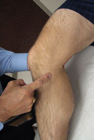
Physical examination for posterolateral knee injury and associated varus laxity
Since most posterolateral corner injuries occur as combined injuries, associated anterior cruciate ligament and posterior cruciate ligament pathology should be looked for.[10][Figure caption and citation for the preceding image starts]: Palpation of the biceps femoris bursaeFrom the personal library of Dr LaPrade; used with permission [Citation ends].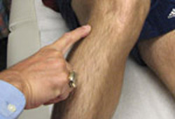
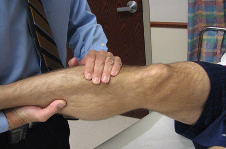
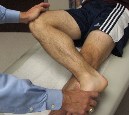
Assessment of varus laxity
This is assessed in a similar manner to valgus laxity. This test looks for an increase in lateral joint line opening. In the evaluation of lateral-sided knee injuries, the contralateral normal knee is also assessed. This is because most patients have a physiologically increased amount of varus opening, usually dependent upon the natural recurvatum of their knee. Thus, it is not possible to determine how much pathology may be present without comparing it with the normal knee.
The thigh is kept stabilised on the examining table while the leg is placed over the foot of the bed. One hand should be on the patient's foot/ankle. This hand is then used to apply the varus force to see how much lateral joint line opening there may be. The other hand should have the fingers directly over the joint line to feel how much joint line opening occurs.[10][15][Figure caption and citation for the preceding image starts]: Varus stress testFrom the personal library of Dr LaPrade; used with permission [Citation ends].
An increased amount of varus opening in extension is usually indicative of a combined posterolateral corner injury and either an anterior cruciate ligament and/or a posterior cruciate ligament.
Varus opening at 30° of knee flexion
Due to the important stabilising effect of the fibular (lateral) collateral ligament, an intact structure should not allow for any increase in the amount of lateral joint line opening compared with the normal knee. An increased amount of varus opening at 30° of knee flexion is therefore indicative of an isolated or combined posterolateral corner injury.
It is important to assess whether there is an actual true opening of the joint or whether there is pseudolaxity present due to lateral compartment arthritis. This can be assessed at the same time as determining how much gapping is present at the joint.
Pitfalls in assessing for varus instability
In patients with open growth plates, a growth plate (physeal) fracture may mimic increased varus joint line opening. Therefore, it is important to utilise stress x-rays in this population.[6]
In older patients with seemingly increased amounts of joint line gapping, there may be associated fractures around the knee, including tibial plateau fractures and distal femoral or supracondylar femur fractures. Patients may have more pain and increased amounts of swelling in these circumstances.
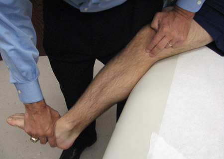
Posterolateral drawer test
The posterolateral drawer test is performed in a similar manner to the posterior drawer test, except that the foot is externally rotated to approximately 15°.
The knee is flexed between 80° and 90°, the foot is externally rotated to approximately 15°, and a posterolateral rotation force is applied to the knee. The amount of posterolateral rotation of the tibia on the femur is observed and the relative subluxation that occurs is noted.[9][10][15] This movement can best be observed by noting how much the tibial tubercle externally rotates in relation to the femur. Due to significant physiological laxity among patients, it is important to grade the increase in posterolateral rotation compared with the normal contralateral knee.
In order to best determine the amount of posterolateral rotation that occurs with the tibia on the femur, it is often useful to sit directly on the patient's foot while applying the coupled posterior drawer and external rotation force. This can be differentiated from a positive anteromedial drawer test, where the tibia rotates anteromedially, which would indicate an MCL and posteromedial joint injury.
The posterolateral drawer test should be differentiated from the posterior drawer test in neutral rotation, because the posterolateral drawer test may be positive even in the face of an intact posterior cruciate ligament.
Increases in the posterolateral drawer test can be due to an isolated posterolateral corner injury, a combined posterolateral corner injury and PCL injury, or, possibly, a knee dislocation. [Figure caption and citation for the preceding image starts]: Posterolateral drawer testFrom the personal library of Dr LaPrade; used with permission [Citation ends].
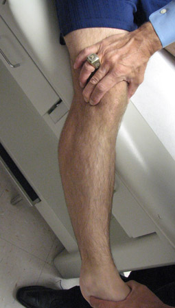
 [Figure caption and citation for the preceding image starts]: Palpation of tibial tubercleFrom the personal library of Dr LaPrade; used with permission [Citation ends].
Pitfalls in assessment of the posterolateral drawer test
Common pitfalls in assessing the posterolateral drawer test are misinterpretation of either the location of the external rotation or the amount of increased posterolateral rotation that is occurring.
Patients who have normal increased recurvatum of their knee will have some increase in their posterolateral drawer test physiologically.[16] In these patients, if the contralateral knee is not assessed, there may appear to be a significant increase in posterolateral rotation on this test.
Posteromedial knee and MCL injury, in which there is an increased amount of anteromedial rotation of the tibia on the femur, may be incorrectly interpreted as being posterolateral rotation.
These differences can be very subtle, but need to be assessed. Other tests, such as varus or valgus stress testing at 30° of knee flexion, may help differentiate if this is a posterolateral corner or, possibly, a medial-sided knee injury.
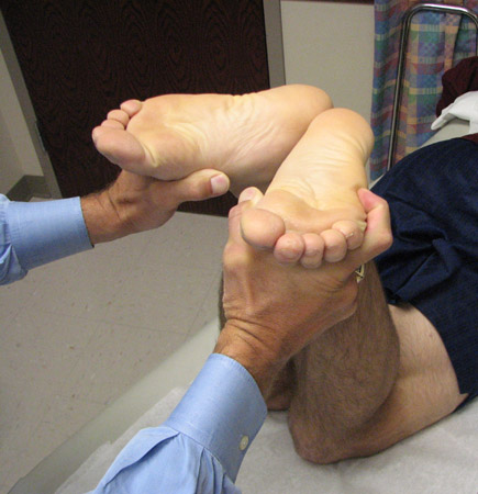
Dial test at 30° in knee flexion
Assesses the amount of external rotation of the tibia on the femur. It can be performed with the patient prone or supine, which makes it useful for patients who have a chronic rather than an acute knee injury, due to their ability to lie prone. Both feet are externally rotated to determine how much increased external rotation of the foot and ankle/foot occurs, compared with the contralateral side.[10][15] Biomechanical laboratory testing has demonstrated that, in the face of a posterolateral corner injury, the knee will externally rotate approximately 15° more than the contralateral normal side.[17] This test makes it easier to discern between the normal and injured knees in patients who are 'tight' and do not have a lot of physiological laxity. In those patients who have increased physiological laxity where their tibial tubercle does rotate normally quite often, it is useful to place the patient in the prone position and assess for the amount of increased external rotation. [Figure caption and citation for the preceding image starts]: Dial test at 30° knee flexion in prone positionFrom the personal library of Dr LaPrade; used with permission [Citation ends].
 [Figure caption and citation for the preceding image starts]: Dial test at 30° knee flexion in supine positionFrom the personal library of Dr LaPrade; used with permission [Citation ends].
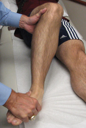
Dial test at 90° in knee flexion
Similar to the dial test at 30°. Can be performed both prone and supine.
When a patient has an isolated posterolateral corner injury, the amount of external rotation of the injured side is approximately 5° more than the contralateral normal knee.
In patients who have a combined posterior cruciate ligament and posterolateral corner injury, or a combined anterior cruciate ligament and posterolateral corner injury, there is an increase in external rotation compared with the uninjured side of approximately 10° to 15°. Therefore, the amount of external rotation that occurs is similar in this injury pattern to the amount of external rotation that is seen at 15° of knee flexion.[10][15][17][Figure caption and citation for the preceding image starts]: Dial test at 90° knee flexion in prone positionFrom the personal library of Dr LaPrade; used with permission [Citation ends].
 [Figure caption and citation for the preceding image starts]: Dial test at 90° knee flexion in supine positionFrom the personal library of Dr LaPrade; used with permission [Citation ends].
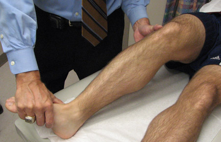
Pitfall in dial test at 90°
An increase in external rotation of the knee can occur when there is a combined MCL and posterior oblique ligament injury to the knee. Therefore, when the dial test is performed prone at 90° of knee flexion, it is possible to mistakenly identify an increase in external rotation at 90° as being due to a posterolateral corner injury when there is in fact a posteromedial corner injury.
It is important to differentiate between posteromedial and posterolateral corner injuries in terms of the amount of external rotation that can occur in the knee.
Reverse pivot shift test
This test is similar to a dynamic posterolateral drawer test. The knee is flexed to approximately 70° to 80°. While cradling the leg with one hand, an external rotation force is applied to the foot and ankle. A positive reverse pivot shift test will result in posterolateral rotation of the tibia on the femur. The knee is then slowly extended and a reduction 'clunk' of the tibia on the femur, where the iliotibial band and popliteus complex reduce the tibia back to its more normal position, is assessed for.[10][15][16][Figure caption and citation for the preceding image starts]: Reverse pivot shift testFrom the personal library of Dr LaPrade; used with permission [Citation ends].
Pitfalls of the reverse pivot shift test interpretation
The reverse pivot shift test has the most false positive results of any clinical knee test. It has been demonstrated that, under anaesthesia, 35% of normal knees have a positive reverse pivot shift test.[16] In the majority of these knees, there is increased physiological laxity, as demonstrated by an increase in genu recurvatum (hyperextension of the knees), as well as increased physiological varus opening of the knee at 30° of knee flexion. Therefore, in the event of a positive reverse pivot shift test, it is very important to differentiate a pathological state versus normal physiological laxity.
In addition, it is possible to incorrectly interpret a positive reverse pivot shift test as a positive pivot shift test. These 2 tests can be differentiated by looking at the starting point of the tibia on the femur and observing how it subluxes. The reverse pivot shift test is different from the pivot shift test in that it starts with the tibia subluxed in flexion rather than in extension, and the tibia is located posterolaterally on the femur rather than anterolaterally.
The figure-4 test
Performed by placing the patient's foot over the contralateral knee. A positive test is indicated by lateral knee pain and suggests a possible tear of the popliteomeniscal fascicles to the lateral meniscus.[18][Figure caption and citation for the preceding image starts]: Figure 4 testFrom the personal library of Dr LaPrade; used with permission [Citation ends].
Gait observations
In patients who have acute posterolateral corner injuries with minimal discomfort and in patients with chronic posterolateral corner knee injuries, especially those with a varus alignment of their lower extremity, ambulation is possible with a varus thrust gait. The mechanism for this gait pattern occurs as the foot strikes the ground. Due to the incompetent posterolateral corner structures there is gapping of the lateral compartment of the knee, which is seen as an obvious thrust of the knee.[6] As the patient shifts his or her weight with gait, and as a result of the dynamic effect of the biceps tendon and the posterolateral corner of the knee, the knee will then close up the lateral compartment gapping. This is observed as a dynamic thrusting of the knee with gait.
Pitfalls in assessment of varus thrust gait
A varus thrust gait pattern due to a posterolateral corner injury is not much different from that observed in a patient who has significant medial compartment arthritis. Therefore, it is important to differentiate between patients who have a varus thrust gait due to medial compartment pseudolaxity versus those who have this gait pattern due to incompetent posterolateral corner structures, resulting in lateral compartment gapping. The physical exam can differentiate between these injury patterns by palpating for lateral compartment opening versus medial compartment collapse, at 30° of knee flexion, during application of a varus stress. Plain anterior-posterior (AP) standing x-rays (where significant medial compartment arthritis is looked for) or bilateral varus stress x-rays (where the amount of lateral compartment gapping that occurs on the injured compared with the normal contralateral side is looked for) can also assist in differentiating between these injury patterns.
Physical examination for posterior cruciate ligament injury
Many symptomatic posterior cruciate ligament injuries are combined with posterolateral corner injuries, and should also be assessed for.
Posterior drawer test
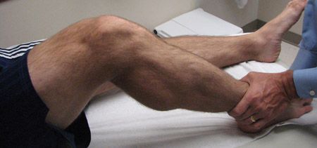
This test is performed with the knee flexed to approximately 80° to 90° and with the foot in neutral rotation. The patient should be sufficiently relaxed such that the hamstrings at the posterior aspect of the knee are completely relaxed. A straight posterior force is then applied through the tibia, and the amount of increase in posterior translation compared with the contralateral knee is assessed. A normal knee will not have any increase in posterior translation.[19][Figure caption and citation for the preceding image starts]: Posterior drawer testFrom the personal library of Dr LaPrade; used with permission [Citation ends].
The posterior drawer test is graded according to how much increased translation there is of the tibia in relation to the distal femur. Grade 1 is an increase in posterior translation, but where the tibia is still anterior to the distal femur. Grade 2 is when the posterior translation of the tibia is such that it is directly in line with the femoral condyles. In those cases where the tibia is subluxed posterior to the anterior edge of the femoral condyles, this is a grade 3 PCL tear and is usually indicative of a combined posterolateral or posteromedial knee injury with the PCL tear.[6]
Pitfalls to performing the posterior drawer test
In patients who cannot relax enough or who may have an injury to the posterior horn of their menisci, any increase in anterior translation may be incorrectly assessed to be an increase in posterior translation.
Posterior sag sign
In this test, both knees are flexed to approximately 90° with the feet resting against the table. In knees that have a posterior cruciate ligament injury, gravity will result in the knee subluxing posteriorly. In thinner knees, this is more obvious. This test facilitates observation of where the tibia sits in relation to the femur. [Figure caption and citation for the preceding image starts]: Posterior sag signFrom the personal library of Dr LaPrade; used with permission [Citation ends].
Quadriceps active test
The patient actively translates the posteriorly subluxed tibia on the femur anteriorly by firing the quadriceps. This is performed with the patient's knee flexed to 90° and the foot on the examining table. The foot is then held and the patient is asked to try to straighten out the knee.
By firing the quadriceps mechanism, a force is transmitted through the patella and patellar tendon on the anterior aspect of the tibia. If the tibia is subluxed posteriorly, the active force of the quadriceps mechanism will reduce the tibia back into its more normal position.[19] In cases where it is difficult to get the patient to relax, it is possible to attempt to push the tibia posteriorly, similar to a posterior drawer, and then have the patient fire the quadriceps mechanism to verify that it is subluxed posteriorly. [Figure caption and citation for the preceding image starts]: Quadriceps active testFrom the personal library of Dr LaPrade; used with permission [Citation ends].
This test is especially useful in patients who may have subtle findings, where it is difficult to determine if they have an ACL or PCL tear. The quadriceps active test will not be positive in the face of a normal posterior cruciate ligament. In patients who may have a multi-ligament injury to their knee and where it is difficult to determine the severity of a potential posterior cruciate ligament injury because of the increased motion of the knee, the quadriceps active test may help to determine if there is a significant PCL injury.
Physical examination for meniscal injuries
Assessment for meniscal tears
This is commonly performed at the same time that medial or lateral joint line opening at 30° in knee flexion is assessed. Patients who have significant trauma may have significant displacement of the meniscus in the joint. These patients may have a lack of full flexion and extension, due either to a bucket handle tear of the posterior aspect of the meniscus anteriorly or to a significant flap tear posteriorly, which can considerably limit their motion. In addition, patients will commonly have joint line crepitation and pain directly over the joint line.[6][20][21][Figure caption and citation for the preceding image starts]: Valgus stress testFrom the personal library of Dr LaPrade; used with permission [Citation ends].
[Figure caption and citation for the preceding image starts]: Varus stress testFrom the personal library of Dr LaPrade; used with permission [Citation ends].Another useful meniscal assessment test is determining if the patient has pain with maximal knee flexion. This test has been shown to be positive in patients who have posterior horn meniscal tears. It is recommended to ask patients who experience pain during deep flexion if the pain is located over the anterior aspect of the knee (which would be more of a patellofemoral source of pain) or posteriorly. One of the most common causes of the posterior knee pain is a meniscus tear; however, patients may have pain from other posterior knee pathology (e.g., a Baker's cyst).
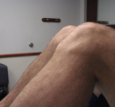
In the assessment of bucket handle tears, it is important to recognise that these do not usually reduce. Therefore, it is appropriate to obtain an MRI scan to assess the extent of the tear before attempting a reduction.[22][23][Figure caption and citation for the preceding image starts]: Deep flexion of the kneeFrom the personal library of Dr LaPrade; used with permission [Citation ends].
Imaging
X-rays
These are usually the first imaging modality following acute trauma to the knee.[24] A knee x-ray series is indicated for people who have sustained a knee injury and demonstrate any of the following:[25]
Age 55 years or older
Isolated tenderness of patella (no bone tenderness of knee other than patella)
Tenderness of head of fibula
Inability to flex to 90°
Inability to bear weight, both immediately and in the emergency department, for 4 steps.
A patellar sunrise x-ray is the most useful if a patellofemoral joint injury is suspected, whereas a standing anterior-posterior (AP) is the first x-ray performed if an anterior cruciate ligament injury is the most likely diagnosis. The reliability and validity of radiologic assessment for patellar instability has been reviewed, but further studies are required.[26]
CT scans
Commonly used to assess for significant bony pathology.[24] This includes the assessment of fractures, tumors, and postsurgical assessments where an MRI may not be possible (patients with pacemakers, significant metal around the knee). Thin slice images (2 mm or less) are recommended to assess the bony architecture of the knee.
Bone scan
This is usually obtained to look for tumors, healing issues after surgery, stress fractures, or other areas of high bone turnover. Thus, 3-phase bone scans with pinholes to better localize the knee architecture are most commonly obtained.
MRI scans of the knee
Useful for determining a diagnosis when there is doubt. MRI scans. They are particularly useful for diagnosis of acute injuries, where bleeding and edema are noted by increased signal uptake on the MRI scans.[22][23] High-field (1.5- or 3-tesla) MRI images are recommended to obtain the greatest detail.
Arteriography
In certain instances (e.g., joint dislocation) arteriography may be helpful to identify deviation or disruption of the vasculature consistent with severe knee pathology.
Invasive testing
Aspiration
Performed if there is an acute effusion. Analysis of a knee aspiration should include aerobic and anaerobic cultures, a cell count and differential of the aspirated fluid, analysis for crystals, and a Gram stain. In general, bloody effusions are more consistent with trauma or tumor (pigmented villonodular synovitis), while nonbloody effusions can be associated with infection (the normal viscosity of the synovial fluid is significantly decreased) or inflammatory arthritis.
Arthroscopy
This is carried out if a meniscal, cartilage, or ligament injury is suspected and the diagnosis needs to be confirmed. Arthroscopy can also be used for treatment of these injuries.
Use of this content is subject to our disclaimer