Images and videos
Images


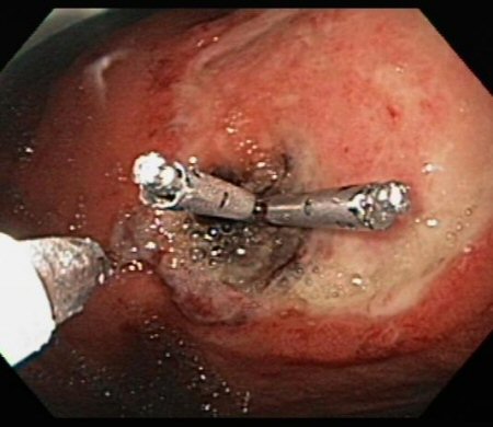
Assessment of upper gastrointestinal bleeding
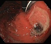
Large gastric ulcer after the endoscopic placement of two mechanical clips to provide haemostasis and prevent rebleeding
From the collection of Douglas G. Adler, MD
See this image in context in the following section/s:
Assessment of upper gastrointestinal bleeding
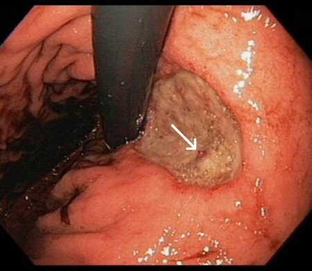
Large gastric ulcer along the lesser curvature with a visible vessel in the ulcer bed (arrow)
From the collection of Douglas G. Adler, MD
See this image in context in the following section/s:
Assessment of upper gastrointestinal bleeding
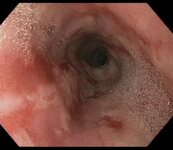
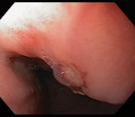
Ulcer in the mid-oesophagus with a visible vessel
From the collection of Douglas G. Adler, MD
See this image in context in the following section/s:
Assessment of upper gastrointestinal bleeding
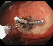
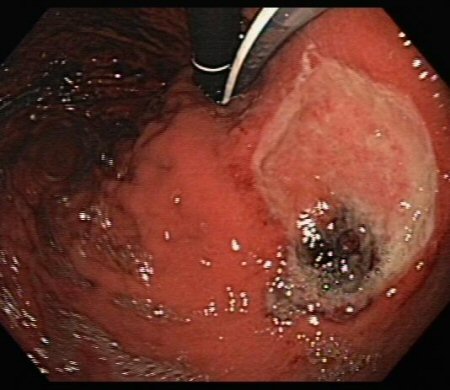
Large gastric ulcer with large, protuberant visible vessel
From the collection of Douglas G. Adler, MD
See this image in context in the following section/s:
Assessment of upper gastrointestinal bleeding
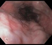
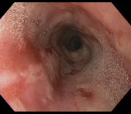
Moderate to severe oesophagitis with multiple linear, clean-based oesophageal ulcers
From the collection of Douglas G. Adler, MD
See this image in context in the following section/s:
Assessment of upper gastrointestinal bleeding
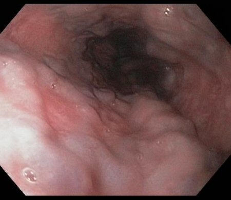
Grade II oesophageal varices in a patient with portal hypertension
From the collection of Douglas G. Adler, MD
See this image in context in the following section/s:
Videos
 Venepuncture and phlebotomy animated demonstration
How to take a venous blood sample from the antecubital fossa using a vacuum needle.
Use of this content is subject to our disclaimer