Images and videos
Images

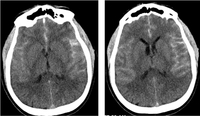
Subarachnoid haemorrhage
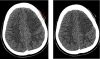
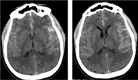
CT brain showing subarachnoid haemorrhage from a ruptured posterior cerebral artery aneurysm (1 of 2)
Courtesy of Dr Salah Keyrouz; used with permission
See this image in context in the following section/s:
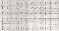
Subarachnoid haemorrhage
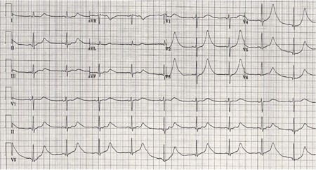
Same patient, 24 hours later; note normalisation of T waves (2 of 2)
Courtesy of Dr Salah Keyrouz; used with permission
See this image in context in the following section/s:
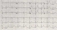
Subarachnoid haemorrhage
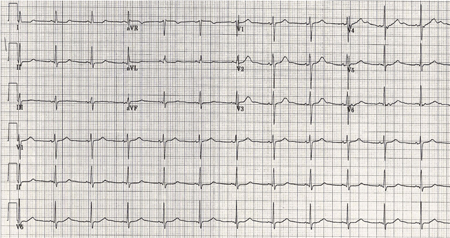
ECG done on admission of a patient with subarachnoid haemorrhage; note peaked, tall T waves (1 of 2)
Courtesy of Dr Salah Keyrouz; used with permission
See this image in context in the following section/s:
Subarachnoid haemorrhage
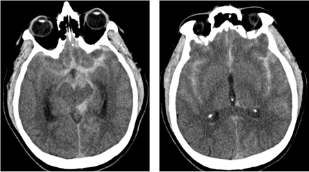
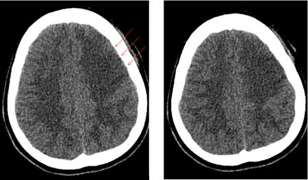
Left frontal infarct (arrows) in a patient with subarachnoid haemorrhage-related vasospasm
Courtesy of Dr Salah Keyrouz; used with permission
See this image in context in the following section/s:
Subarachnoid haemorrhage
CT brain showing subarachnoid haemorrhage from a ruptured posterior cerebral artery aneurysm (2 of 2)
Courtesy of Dr Salah Keyrouz; used with permission
See this image in context in the following section/s:
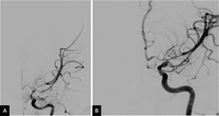
Subarachnoid haemorrhage
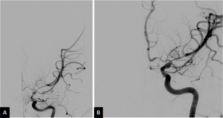
Severe vasospasm of distal left internal carotid artery and proximal middle and anterior cerebral arteries before (A) and after (B) intra-arterial infusion of nicardipine and transluminal balloon angioplasty
Courtesy of Dr Salah Keyrouz; used with permission
See this image in context in the following section/s:
Videos
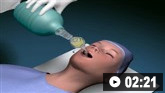
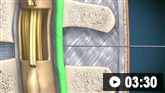 Diagnostic lumbar puncture in adults: animated demonstration
How to perform a diagnostic lumbar puncture in adults. Includes a discussion of patient positioning, choice of needle, and measurement of opening and closing pressure.
Use of this content is subject to our disclaimer