Aetiology
Several genotypes of Giardia duodenalis have been discovered but only a few are host-specific to humans.[21]
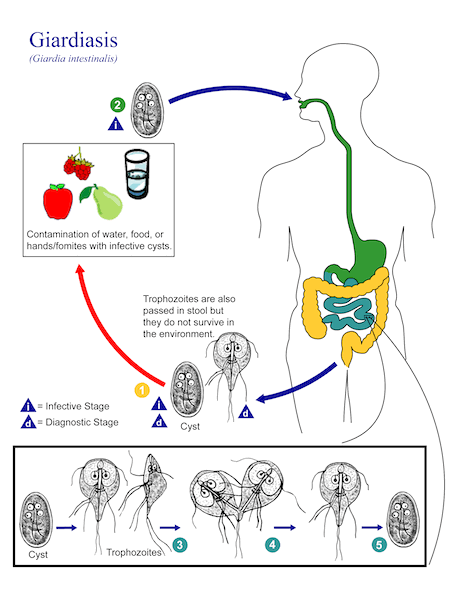
There are two morphological forms of G duodenalis: cysts and trophozoites. Infection requires oral ingestion of cysts (fewer than 10 cysts are sufficient to cause infection). The incubation period ranges from 1-25 days (average 7 days).[7][22][23]
After ingestion of cysts, excystation occurs in the proximal small bowel where trophozoites are released. These attach to the mucosal surface of the duodenum and jejunum by using an adhesive disk on their ventral surface. Trophozoites are converted to the infectious cyst form in the small intestine.[19][Figure caption and citation for the preceding image starts]: Scanning electron micrograph of Giardia trophozoite on the mucosal surface of the intestine. The ventral adhesive disc, which facilitates adherence to the intestinal surface, can be seen on the underside of the organismCourtesy of Dr Stan Erlandsen; CDC Public Health Image Library (PHIL) [Citation ends].
Acquisition of Giardia is by ingestion of water (or food) contaminated by cysts and by person-to-person spread through the faecal-oral route.[19] Being in contact with children in nappies; drinking water from, or swimming in a, river, lake, or stream (e.g., campers, hunters, backpackers); international travel; tourism to endemic areas; oral-anal sexual contact; and eating lettuce are risk factors for developing giardiasis.[16][17][18][19][24][25]
Common variable late-onset hypogammaglobulinaemia has been associated with a high frequency of giardiasis.[26] Recent antibiotic use and history of gastrointestinal disorder are emerging risk factors for sporadic infections.[18] Sporadic cases can occur without an identifiable risk factor.[Figure caption and citation for the preceding image starts]: Illustration of the life cycle of Giardia duodenalis (formerly known as Giardia lamblia or Giardia intestinalis), the causal agent of giardiasisCourtesy of Alexander J. da Silva, Melanie Moser; CDC Public Health Image Library (PHIL) [Citation ends].
Pathophysiology
Pathogenesis of giardiasis is not completely understood and several hypotheses exist. Theories include direct damage to the brush border intestinal mucosa either by trophozoites or by host immune-response activation. Some animal models report reduced activity of digestive enzymes in the gut and/or impaired gut barrier function.[4][27][28] Certain bile acids present in the small intestine promote Giardia survival.[29] The characteristic greasy, foul-smelling stools indicative of malabsorption may be due to bile acids being taken up by the organism, but are not a frequent presenting symptom.
Giardia infection is associated with disruptions in gut bacterial communities. In experimental models, these disruptions contribute to pathogenesis through either gut dysfunction and/or perturbed metabolism that may contribute to impaired growth potential.[4][30][31][32][33]
Giardia disrupts tight junctions in human intestinal monolayers, which increases epithelial permeability and can result in nutrient deficiency.[34][35] and results in fat malabsorption. Weight loss and malabsorption of fat-soluble vitamins, lactose, and vitamin B12 are commonly seen in chronic giardiasis. Children can present with faltering growth, wasting, or stunting.
Giardia and infection-induced gut inflammation can induce programmed cell death (apoptosis).[36][37] This could cause malabsorption of sodium, water and glucose and reduced disaccharidase activity, due to loss of absorptive surface area of the epithelium.[38][39][40][41]
Use of this content is subject to our disclaimer