Approach
Early diagnosis of TSS is essential, as serious life-threatening infections can develop within 24 to 72 hours. Initial signs and symptoms are usually non-specific: for example, pain, fever, malaise, gastrointestinal symptoms such as vomiting and diarrhoea, and muscle aches. The clinical course of TSS is often precipitous, and requires a high index of suspicion, prompt diagnosis, and early treatment in an intensive care unit.
Choice of antibiotics in treatment of TSS is aided by differentiating between streptococcal and staphylococcal disease, although the two have many similar history and examination factors and test results. Microbiological cultures are not always the best differentiating points because of low likelihood of producing a positive culture.
Clinical assessment
Streptococcal TSS should be considered in a healthy patient from the community who presents with shock, severe pain, and fever and has a recent history of trauma. TSS should also be considered in patients with a relatively unimpressive focus of infection but evidence of septic shock. In many cases of streptococcal TSS, no site of entry is found, or it is subtle.[41] Invasive streptococcal infections include bacteraemia, cellulitis, meningitis, pneumonia, empyema, peritonitis, septic arthritis, puerperal sepsis, burn wound sepsis, necrotising fasciitis, or gangrenous myositis.[9] Serious bacterial infections caused by group A streptococci infections are increasing in frequency as a complication of varicella.[10][11]
Signs and symptoms
The most common initial presentation of TSS from a group A streptococcal invasive infection is abrupt onset of severe diffuse or localised pain, which often precedes the physical findings of a soft-tissue infection.[63] Extremity pain is the most common complaint, but the pain may mimic peritonitis, PID, pneumonia, MI, pericarditis, or cholecystitis.[63][81][82] Eighty percent have clinical signs of a soft-tissue infection, with localised swelling or erythema with subsequent ecchymoses and skin sloughing progressing to necrotising fasciitis or myositis in 70% of cases.[41] Gangrene may also be present.
Staphylococcal TSS should be considered in menstruating patients who have used tampons. However, suspicion should not be limited to only these patients, as non-menstruating patients can develop staphylococcal TSS.[21][22]
In both streptococcal and staphylococcal infections, fever is the most common presenting sign, although hypothermia can be associated with shock.[63] Non-specific signs and symptoms on presentation, such as fever, chills, nausea, vomiting, diarrhoea, and myalgias, are present in 20% of patients, typically associated with toxin production.[68] A diffuse, erythematous rash develops in 10% of patients with streptococcal TSS. In staphylococcal disease, the maculopapular rash desquamates in 1 to 2 weeks and is often seen initially on the palms and soles. Half of all patients are normotensive on admission, but hypotension and shock will usually develop in the following 4 to 8 hours.[63] Other signs and symptoms of shock and hypoperfusion can be present, such as mental status changes and oliguria.
Myocarditis, peritonitis, and endophthalmitis may be present in both streptococcal and staphylococcal infections. Myocarditis can manifest as chest pain, dyspnoea, orthopnoea, syncope, fatigue, and palpitations. Findings can include signs of cardiac failure, tachycardias, or arrhythmias. Patients with peritonitis may present with severe abdominal pain with rebound tenderness and guarding on abdominal examination.[Figure caption and citation for the preceding image starts]: Subtle desquamation of the finger tips of the left hand caused by toxic shock syndromeFrom the CDC and the Public Health Image Library [Citation ends].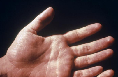 [Figure caption and citation for the preceding image starts]: Patient with facial erythematous rash due to toxic shock syndromeFrom the CDC and the Public Health Image Library [Citation ends].
 [Figure caption and citation for the preceding image starts]: Rash and subcutaneous oedema of the right hand due to toxic shock syndromeFrom the CDC and the Public Health Image Library [Citation ends].
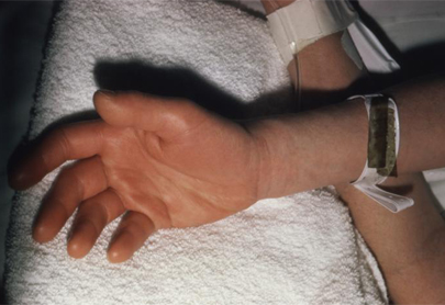 [Figure caption and citation for the preceding image starts]: Patient displaying a morbilliform rash (resembling measles) resulting from toxic shock syndrome, 3 to 5 days after onsetFrom the CDC and the Public Health Image Library [Citation ends].
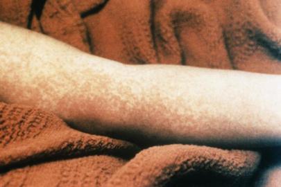 [Figure caption and citation for the preceding image starts]: Marked desquamation of the left palm due to toxic shock syndrome, which develops late in the diseaseFrom the CDC and the Public Health Image Library [Citation ends].
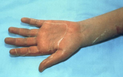
Risk stratification: identifying patients at risk of deterioration due to organ dysfunction
Early identification of sepsis relies on the systematic evaluation of patients presenting with presumed infection to identify those at risk of deterioration due to organ dysfunction.
Several approaches have been proposed for the quick and pragmatic evaluation of risk of deterioration in everyday clinical practice without the need to await laboratory investigations. Recommendations vary among hospitals and countries. Further research is required to determine the optimal approach, which includes early warning scores (e.g., the National Early Warning Score 2 [NEWS2] developed in the UK or the Modified Early Warning Score [MEWS]), or the use of a risk stratification system as recommended by various guideline groups in the US and UK.[83][84][85][86][87] These scores typically incorporate physiological variables such as heart rate and blood pressure, and they can easily be calculated at the bedside. None have been evaluated specifically for TSS.
Investigations
The following investigations should be performed on all patients:
Microscopy and culture on normally sterile sites (blood, cerebrospinal fluid, pleural or peritoneal fluid, tissue, or throat) may be positive for group A streptococcus or Staphylococcus aureus. However, of patients with streptococcal TSS, 60% have positive blood cultures, and of patients with staphylococcal TSS, <5% have positive blood cultures[41]
FBC and differential show leukocytosis, anaemia, and thrombocytopenia[63]
Renal function: elevated urea, creatinine, and haemoglobinuria are signs of renal failure. Precedes hypotension in 40% to 50% of patients with streptococcal disease[44]
Liver function: elevation of bilirubin or transaminases more than twice the normal upper limit[2]
Sodium, phosphorus, albumin, and calcium: commonly low on admission with streptococcal disease and throughout the clinical course
Lactic acid: elevated in severe sepsis and septic shock
Elevated creatine kinase (CK) suggests necrotising fasciitis or myositis. CK may also be elevated in staphylococcal TSS
Coagulation profile: shows increased prothrombin and partial thromboplastin times in staphylococcal disease in conjunction with disseminated intravascular coagulation (DIC); fibrinogen: may be low in the setting of DIC.
Acute and convalescent staphylococcal antibody testing: positive results may support the diagnosis of staphylococcal TSS
Specific testing of streptococcal exotoxin serotypes is possible, but these tests are not routinely available in most hospitals.
Imaging
Chest x-ray should be performed after initial blood and culture work-up and may show diffuse bilateral interstitial and alveolar infiltrates consistent with acute respiratory distress syndrome in both streptococcal and staphylococcal disease.
Clinicians should have a low threshold for obtaining definitive imaging to determine the source of infection, because a focus of infection can be subtle in TSS.
Use of this content is subject to our disclaimer