The majority of patients with acute HEV infection are asymptomatic. In patients who develop an acute hepatitis (possibly less than 5% of all cases of HEV infection), the history should look for risk factors for potential exposure to HEV - most commonly, living in or recent travel to endemic areas such as Africa or Asia (transmission via the faecal-oral route due to genotype 1 or 2 infection), or via consumption of raw or undercooked pork, wild boar, or deer (food-borne/zoonotic transmission due to genotype 3 or 4), particularly in Europe.[1]Centers for Disease Control and Prevention. Viral hepatitis: hepatitis E. Jun 2020 [internet publication].
https://www.cdc.gov/hepatitis/hev/index.htm
[2]European Association for the Study of the Liver. EASL clinical practice guidelines on hepatitis E virus infection. J Hepatol. 2018 Jun;68(6):1256-71.
https://www.journal-of-hepatology.eu/article/S0168-8278(18)30155-7/fulltext
http://www.ncbi.nlm.nih.gov/pubmed/29609832?tool=bestpractice.com
Many patients in developed countries will not have a readily identifiable risk factor so, in these regions, any patient with an otherwise unexplained acute hepatitis could have HEV infection.[2]European Association for the Study of the Liver. EASL clinical practice guidelines on hepatitis E virus infection. J Hepatol. 2018 Jun;68(6):1256-71.
https://www.journal-of-hepatology.eu/article/S0168-8278(18)30155-7/fulltext
http://www.ncbi.nlm.nih.gov/pubmed/29609832?tool=bestpractice.com
If symptoms do occur with acute HEV infection, they are usually non-specific and may be indistinguishable from other types of acute hepatitis.
Symptomatic infection is most common in those aged 44 years and younger in developing countries, where infection is usually acquired via the faeco-oral route.[1]Centers for Disease Control and Prevention. Viral hepatitis: hepatitis E. Jun 2020 [internet publication].
https://www.cdc.gov/hepatitis/hev/index.htm
In developed countries, symptomatic illness is most commonly seen in older men, which may indicate the presence of underlying liver disease.[11]Kumar Acharya S, Kumar Sharma P, Singh R, et al. Hepatitis E virus (HEV) infection in patients with cirrhosis is associated with rapid decompensation and death. J Hepatol. 2007 Mar;46(3):387-94.
http://www.ncbi.nlm.nih.gov/pubmed/17125878?tool=bestpractice.com
Progression to acute liver failure is rare, and is more of a concern in patients with existing chronic liver disease or patients who are pregnant; it is therefore important to have a high index of suspicion for HEV infection in pregnant patients who present with an acute hepatitis in endemic areas.[2]European Association for the Study of the Liver. EASL clinical practice guidelines on hepatitis E virus infection. J Hepatol. 2018 Jun;68(6):1256-71.
https://www.journal-of-hepatology.eu/article/S0168-8278(18)30155-7/fulltext
http://www.ncbi.nlm.nih.gov/pubmed/29609832?tool=bestpractice.com
HEV infection acquired via the faeco-oral route is associated with high morbidity and mortality in pregnant patients.[1]Centers for Disease Control and Prevention. Viral hepatitis: hepatitis E. Jun 2020 [internet publication].
https://www.cdc.gov/hepatitis/hev/index.htm
[2]European Association for the Study of the Liver. EASL clinical practice guidelines on hepatitis E virus infection. J Hepatol. 2018 Jun;68(6):1256-71.
https://www.journal-of-hepatology.eu/article/S0168-8278(18)30155-7/fulltext
http://www.ncbi.nlm.nih.gov/pubmed/29609832?tool=bestpractice.com
[17]Kamar N, Bendall R, Legrand-Abravanel F, et al. Hepatitis E. Lancet. 2012 Jun 30;379(9835):2477-88.
http://www.ncbi.nlm.nih.gov/pubmed/22549046?tool=bestpractice.com
Extrahepatic manifestations of HEV infection can occur after acute infection and with chronic disease. Most significantly, there are a myriad of neurological syndromes associated with HEV; an acute hepatitis with neurological symptoms should raise suspicion for HEV infection.
All patients who present with signs and symptoms of acute hepatitis or acute liver failure, immunosuppressed patients, and patients with underlying liver disease with unexplained elevation of liver function tests should be tested for HEV infection.[2]European Association for the Study of the Liver. EASL clinical practice guidelines on hepatitis E virus infection. J Hepatol. 2018 Jun;68(6):1256-71.
https://www.journal-of-hepatology.eu/article/S0168-8278(18)30155-7/fulltext
http://www.ncbi.nlm.nih.gov/pubmed/29609832?tool=bestpractice.com
The incubation period after exposure for HEV is 2-8 weeks; HEV RNA can be detected in stool about 3 weeks after infection and can last for approximately 6 weeks.[46]Kamani L, Padhani ZA, Das JK. Hepatitis E: genotypes, strategies to prevent and manage, and the existing knowledge gaps. JGH Open. 2021 Oct;5(10):1127-34.
https://onlinelibrary.wiley.com/doi/10.1002/jgh3.12646
http://www.ncbi.nlm.nih.gov/pubmed/34621997?tool=bestpractice.com
[47]Chandra NS, Sharma A, Malhotra B, et al. Dynamics of HEV viremia, fecal shedding and its relationship with transaminases and antibody response in patients with sporadic acute hepatitis E. Virol J. 2010 Sep 6;7:213.
https://virologyj.biomedcentral.com/articles/10.1186/1743-422X-7-213
http://www.ncbi.nlm.nih.gov/pubmed/20815928?tool=bestpractice.com
Biochemical and serological markers start to increase just prior to symptom onset.[2]European Association for the Study of the Liver. EASL clinical practice guidelines on hepatitis E virus infection. J Hepatol. 2018 Jun;68(6):1256-71.
https://www.journal-of-hepatology.eu/article/S0168-8278(18)30155-7/fulltext
http://www.ncbi.nlm.nih.gov/pubmed/29609832?tool=bestpractice.com
Progression to chronic liver disease is most commonly associated with patients who are immunosuppressed, such as recipients of solid organ transplants, and has been defined as persistence of HEV replication for 3 or more months.[2]European Association for the Study of the Liver. EASL clinical practice guidelines on hepatitis E virus infection. J Hepatol. 2018 Jun;68(6):1256-71.
https://www.journal-of-hepatology.eu/article/S0168-8278(18)30155-7/fulltext
http://www.ncbi.nlm.nih.gov/pubmed/29609832?tool=bestpractice.com
[4]Kamar N, Selves J, Mansuy JM, et al. Hepatitis E virus and chronic hepatitis in organ-transplant recipients. N Engl J Med. 2008 Feb 21;358(8):811-7.
https://www.nejm.org/doi/full/10.1056/NEJMoa0706992
http://www.ncbi.nlm.nih.gov/pubmed/18287603?tool=bestpractice.com
[5]Marion O, Abravanel F, Lhomme S, et al. Hepatitis E in transplantation. Curr Infect Dis Rep. 2016 Mar;18(3):8.
http://www.ncbi.nlm.nih.gov/pubmed/26838163?tool=bestpractice.com
Chronic infection has only been associated with infection with HEV genotypes 3 and 4.[4]Kamar N, Selves J, Mansuy JM, et al. Hepatitis E virus and chronic hepatitis in organ-transplant recipients. N Engl J Med. 2008 Feb 21;358(8):811-7.
https://www.nejm.org/doi/full/10.1056/NEJMoa0706992
http://www.ncbi.nlm.nih.gov/pubmed/18287603?tool=bestpractice.com
[48]Gérolami R, Moal V, Colson P. Chronic hepatitis E with cirrhosis in a kidney-transplant recipient. N Engl J Med. 2008 Feb 21;358(8):859-60.
https://www.nejm.org/doi/full/10.1056/NEJMc0708687
http://www.ncbi.nlm.nih.gov/pubmed/18287615?tool=bestpractice.com
Solid organ transplant recipients infected with HEV have a greater than 50% risk of developing chronic HEV infection, which can progress to cirrhosis within several years.[38]Péron JM. Hepatitis E virus infection and cirrhosis of the liver. Gastroenterol Hepatol (N Y). 2016 Sep;12(9):565-7.
https://www.gastroenterologyandhepatology.net/archives/september-2016/hepatitis-e-virus-infection-and-cirrhosis-of-the-liver
http://www.ncbi.nlm.nih.gov/pubmed/27917096?tool=bestpractice.com
[49]Pischke S, Stiefel P, Franz B, et al. Chronic hepatitis E in heart transplant recipients. Am J Transplant. 2012 Nov;12(11):3128-33.
https://www.amjtransplant.org/article/S1600-6135(22)27702-8/fulltext
http://www.ncbi.nlm.nih.gov/pubmed/22823202?tool=bestpractice.com
Chronic infection has less commonly been associated with other immunosuppressed groups, such as people with primary immunodeficiency, those with HIV who have low CD4 T lymphocyte counts, and those undergoing chemotherapy (especially due to haematological malignancies).[2]European Association for the Study of the Liver. EASL clinical practice guidelines on hepatitis E virus infection. J Hepatol. 2018 Jun;68(6):1256-71.
https://www.journal-of-hepatology.eu/article/S0168-8278(18)30155-7/fulltext
http://www.ncbi.nlm.nih.gov/pubmed/29609832?tool=bestpractice.com
[38]Péron JM. Hepatitis E virus infection and cirrhosis of the liver. Gastroenterol Hepatol (N Y). 2016 Sep;12(9):565-7.
https://www.gastroenterologyandhepatology.net/archives/september-2016/hepatitis-e-virus-infection-and-cirrhosis-of-the-liver
http://www.ncbi.nlm.nih.gov/pubmed/27917096?tool=bestpractice.com
Any cause of acute hepatocellular injury is a differential for acute or chronic HEV infection. For chronic HEV infection, the differential diagnosis includes diseases seen in immunosuppressed patients, such as other viral causes of hepatitis. Several severe diseases seen in pregnancy are also differentials of acute HEV infection in pregnant individuals. See Differentials.
History
Acute HEV infection
If symptoms do occur with acute HEV infection they are usually non-specific, and may include:
Malaise[2]European Association for the Study of the Liver. EASL clinical practice guidelines on hepatitis E virus infection. J Hepatol. 2018 Jun;68(6):1256-71.
https://www.journal-of-hepatology.eu/article/S0168-8278(18)30155-7/fulltext
http://www.ncbi.nlm.nih.gov/pubmed/29609832?tool=bestpractice.com
Fatigue[2]European Association for the Study of the Liver. EASL clinical practice guidelines on hepatitis E virus infection. J Hepatol. 2018 Jun;68(6):1256-71.
https://www.journal-of-hepatology.eu/article/S0168-8278(18)30155-7/fulltext
http://www.ncbi.nlm.nih.gov/pubmed/29609832?tool=bestpractice.com
Anorexia[2]European Association for the Study of the Liver. EASL clinical practice guidelines on hepatitis E virus infection. J Hepatol. 2018 Jun;68(6):1256-71.
https://www.journal-of-hepatology.eu/article/S0168-8278(18)30155-7/fulltext
http://www.ncbi.nlm.nih.gov/pubmed/29609832?tool=bestpractice.com
Nausea[2]European Association for the Study of the Liver. EASL clinical practice guidelines on hepatitis E virus infection. J Hepatol. 2018 Jun;68(6):1256-71.
https://www.journal-of-hepatology.eu/article/S0168-8278(18)30155-7/fulltext
http://www.ncbi.nlm.nih.gov/pubmed/29609832?tool=bestpractice.com
Vomiting[2]European Association for the Study of the Liver. EASL clinical practice guidelines on hepatitis E virus infection. J Hepatol. 2018 Jun;68(6):1256-71.
https://www.journal-of-hepatology.eu/article/S0168-8278(18)30155-7/fulltext
http://www.ncbi.nlm.nih.gov/pubmed/29609832?tool=bestpractice.com
Abdominal pain[2]European Association for the Study of the Liver. EASL clinical practice guidelines on hepatitis E virus infection. J Hepatol. 2018 Jun;68(6):1256-71.
https://www.journal-of-hepatology.eu/article/S0168-8278(18)30155-7/fulltext
http://www.ncbi.nlm.nih.gov/pubmed/29609832?tool=bestpractice.com
Pruritus[2]European Association for the Study of the Liver. EASL clinical practice guidelines on hepatitis E virus infection. J Hepatol. 2018 Jun;68(6):1256-71.
https://www.journal-of-hepatology.eu/article/S0168-8278(18)30155-7/fulltext
http://www.ncbi.nlm.nih.gov/pubmed/29609832?tool=bestpractice.com
Arthralgia/arthritis[2]European Association for the Study of the Liver. EASL clinical practice guidelines on hepatitis E virus infection. J Hepatol. 2018 Jun;68(6):1256-71.
https://www.journal-of-hepatology.eu/article/S0168-8278(18)30155-7/fulltext
http://www.ncbi.nlm.nih.gov/pubmed/29609832?tool=bestpractice.com
Diarrhoea.
Enquire about potential exposure to HEV in patients who present with acute hepatitis, such as travel to or from endemic areas, or consumption of swine or deer, particularly in Europe.[1]Centers for Disease Control and Prevention. Viral hepatitis: hepatitis E. Jun 2020 [internet publication].
https://www.cdc.gov/hepatitis/hev/index.htm
Take a detailed history regarding travel, sources of drinking water, consumption of uncooked or undercooked food, and recent contact with any jaundiced people.[1]Centers for Disease Control and Prevention. Viral hepatitis: hepatitis E. Jun 2020 [internet publication].
https://www.cdc.gov/hepatitis/hev/index.htm
Infection with HEV genotypes 1 and 2 is typically acquired by drinking faecally contaminated water in endemic areas, demonstrated by epidemic peaks during the rainy season.[1]Centers for Disease Control and Prevention. Viral hepatitis: hepatitis E. Jun 2020 [internet publication].
https://www.cdc.gov/hepatitis/hev/index.htm
[7]Labrique AB, Zaman K, Hossain Z, et al. Epidemiology and risk factors of incident hepatitis E virus infections in rural Bangladesh. Am J Epidemiol. 2010 Oct 15;172(8):952-61.
https://academic.oup.com/aje/article/172/8/952/275587
http://www.ncbi.nlm.nih.gov/pubmed/20801864?tool=bestpractice.com
HEV genotypes 3 and 4 are acquired by ingestion of contaminated food or close association with the animal reservoir, such as by farm workers or veterinarians.[19]Meng XJ, Wiseman B, Elvinger F, et al. Prevalence of antibodies to hepatitis E virus in veterinarians working with swine and in normal blood donors in the United States and other countries. J Clin Microbiol. 2002 Jan;40(1):117-22.
https://journals.asm.org/doi/10.1128/jcm.40.1.117-122.2002
http://www.ncbi.nlm.nih.gov/pubmed/11773103?tool=bestpractice.com
[29]Chaussade H, Rigaud E, Allix A, et al. Hepatitis E virus seroprevalence and risk factors for individuals in working contact with animals. J Clin Virol. 2013 Nov;58(3):504-8.
http://www.ncbi.nlm.nih.gov/pubmed/24084601?tool=bestpractice.com
Although much less common, HEV infection can occur after transfusion of blood products or following organ transplantation.[22]Hewitt PE, Ijaz S, Brailsford SR, et al. Hepatitis E virus in blood components: a prevalence and transmission study in southeast England. Lancet. 2014 Nov 15;384(9956):1766-73.
https://www.thelancet.com/journals/lancet/article/PIIS0140-6736(14)61034-5/fulltext
http://www.ncbi.nlm.nih.gov/pubmed/25078306?tool=bestpractice.com
[30]Cheung CKM, Wong SH, Law AWH, et al. Transfusion-transmitted hepatitis E: what we know so far? World J Gastroenterol. 2022 Jan 7;28(1):47-75.
https://www.wjgnet.com/1007-9327/full/v28/i1/47.htm
http://www.ncbi.nlm.nih.gov/pubmed/35125819?tool=bestpractice.com
[31]Slavov SN, Maçonetto JDM, Martinez EZ, et al. Prevalence of hepatitis E virus infection in multiple transfused Brazilian patients with thalassemia and sickle cell disease. J Med Virol. 2019 Sep;91(9):1693-7.
http://www.ncbi.nlm.nih.gov/pubmed/31066064?tool=bestpractice.com
[32]Public Health England. Hepatitis E: symptoms, transmission, treatment and prevention. May 2020 [internet publication].
https://www.gov.uk/government/publications/hepatitis-e-symptoms-transmission-prevention-treatment/hepatitis-e-symptoms-transmission-treatment-and-prevention
[33]Ushiro-Lumb I, Forsythe J, Haywood B, et al. Impact of hepatitis E virus screening in the UK deceased organ donor population. Transpl Int. 2023 Sep 4;36:11673.
https://www.frontierspartnerships.org/articles/10.3389/ti.2023.11673/full
http://www.ncbi.nlm.nih.gov/pubmed/37727381?tool=bestpractice.com
These risks are not applicable universally: for example, in the UK, blood donations are screened for HEV before transfusion.
Vertical transmission can occur, but this is a less common mode of transmission.[34]Kumar RM, Uduman S, Rana S, et al. Sero-prevalence and mother-to-infant transmission of hepatitis E virus among pregnant women in the United Arab Emirates. Eur J Obstet Gynecol Reprod Biol. 2001 Dec 10;100(1):9-15.
http://www.ncbi.nlm.nih.gov/pubmed/11728649?tool=bestpractice.com
People living in crowded camps or temporary housing, such as refugees or people who are internally displaced, or those living in military camps, are at greater risk of contracting HEV infection.[1]Centers for Disease Control and Prevention. Viral hepatitis: hepatitis E. Jun 2020 [internet publication].
https://www.cdc.gov/hepatitis/hev/index.htm
[40]Tsega E, Krawczynski K, Hansson BG, et al. Outbreak of acute hepatitis E virus infection among military personnel in northern Ethiopia. J Med Virol. 1991 Aug;34(4):232-6.
http://www.ncbi.nlm.nih.gov/pubmed/1940876?tool=bestpractice.com
Ask about previous medical history as well as symptoms of extrahepatic manifestations of HEV, which can occur after acute infection and with chronic disease. As there are myriad neurological syndromes associated with HEV infection, an acute hepatitis with neurological symptoms should raise suspicion for the condition. Conditions that have been observed in the context of hepatitis E are numerous, and include:[2]European Association for the Study of the Liver. EASL clinical practice guidelines on hepatitis E virus infection. J Hepatol. 2018 Jun;68(6):1256-71.
https://www.journal-of-hepatology.eu/article/S0168-8278(18)30155-7/fulltext
http://www.ncbi.nlm.nih.gov/pubmed/29609832?tool=bestpractice.com
[9]Pischke S, Hartl J, Pas SD, et al. Hepatitis E virus: infection beyond the liver? J Hepatol. 2017 May;66(5):1082-95.
http://www.ncbi.nlm.nih.gov/pubmed/27913223?tool=bestpractice.com
Neurological
Guillain-Barré syndrome
Neuralgic amyotrophy
Meningitis
Cranial nerve palsies
Renal
Haematological
Cryoglobulinaemia
Thrombocytopenia
Haemolysis
Aplastic anaemia
Other
Acute pancreatitis
Thyroiditis
Myocarditis.
Acute liver failure/acute-on-chronic liver failure
Although the definition of acute liver failure varies globally, the most commonly used definition in the US and Europe is: an illness duration of <26 weeks in a patient with no evidence of prior liver disease or cirrhosis with any degree of mental status alteration (encephalopathy) and coagulopathy (international normalised ratio [INR] ≥1.5).[50]Shingina A, Mukhtar N, Wakim-Fleming J, et al. Acute liver failure guidelines. Am J Gastroenterol. 2023 Jul 1;118(7):1128-53.
https://journals.lww.com/ajg/fulltext/2023/07000/acute_liver_failure_guidelines.14.aspx
http://www.ncbi.nlm.nih.gov/pubmed/37377263?tool=bestpractice.com
Acute liver failure is a rare sequela of acute HEV infection and is more of a concern in patients with existing chronic liver disease (when symptoms occur in a patient with pre-existing liver disease, the term 'acute-on-chronic liver failure' is used).[2]European Association for the Study of the Liver. EASL clinical practice guidelines on hepatitis E virus infection. J Hepatol. 2018 Jun;68(6):1256-71.
https://www.journal-of-hepatology.eu/article/S0168-8278(18)30155-7/fulltext
http://www.ncbi.nlm.nih.gov/pubmed/29609832?tool=bestpractice.com
[11]Kumar Acharya S, Kumar Sharma P, Singh R, et al. Hepatitis E virus (HEV) infection in patients with cirrhosis is associated with rapid decompensation and death. J Hepatol. 2007 Mar;46(3):387-94.
http://www.ncbi.nlm.nih.gov/pubmed/17125878?tool=bestpractice.com
However, it is important to note that acute HEV infection in pregnancy in endemic areas can lead to acute liver failure in upwards of 20% of women.[10]Patra S, Kumar A, Trivedi SS, et al. Maternal and fetal outcomes in pregnant women with acute hepatitis E virus infection. Ann Intern Med. 2007 Jul 3;147(1):28-33.
http://www.ncbi.nlm.nih.gov/pubmed/17606958?tool=bestpractice.com
Patients who progress to acute, or acute-on-chronic, liver failure may present with symptoms including:[51]Trey C, Davidson CS. The management of fulminant hepatic failure. In: Popper H, Shaffner F (eds). Progress in liver diseases. 3rd ed. New York, NY: Grune & Stratton; 1970:282-98.
http://www.ncbi.nlm.nih.gov/pubmed/4908702?tool=bestpractice.com
[52]Gimson AE, O'Grady J, Ede RJ, et al. Late onset hepatic failure: clinical, serological and histological features. Hepatology. 1986 Mar-Apr;6(2):288-94.
https://aasldpubs.onlinelibrary.wiley.com/doi/epdf/10.1002/hep.1840060222
http://www.ncbi.nlm.nih.gov/pubmed/3082735?tool=bestpractice.com
[53]Bernuau J, Rueff B, Benhamou JP. Fulminant and subfulminant liver failure: definitions and causes. Semin Liver Dis. 1986 May;6(2):97-106.
http://www.ncbi.nlm.nih.gov/pubmed/3529410?tool=bestpractice.com
See Acute liver failure.
Chronic HEV infection
Clinical presentation of chronic HEV infection has largely been found in patients with solid organ transplantation, but has also been associated with other immunosuppressed groups, such as individuals with HIV, patients with primary immunodeficiencies, and patients undergoing chemotherapy.[2]European Association for the Study of the Liver. EASL clinical practice guidelines on hepatitis E virus infection. J Hepatol. 2018 Jun;68(6):1256-71.
https://www.journal-of-hepatology.eu/article/S0168-8278(18)30155-7/fulltext
http://www.ncbi.nlm.nih.gov/pubmed/29609832?tool=bestpractice.com
[13]Suneetha PV, Pischke S, Schlaphoff V, et al. Hepatitis E virus (HEV)-specific T-cell responses are associated with control of HEV infection. Hepatology. 2012 Mar;55(3):695-708.
http://www.ncbi.nlm.nih.gov/pubmed/22006345?tool=bestpractice.com
Chronic HEV infection is frequently asymptomatic in people who are immunosuppressed, and is typically identified during investigation following abnormal biochemical liver function tests.
Solid organ transplant recipients infected with HEV have between a 50% and 70% risk of developing chronic HEV infection, which can progress to cirrhosis within several years.[2]European Association for the Study of the Liver. EASL clinical practice guidelines on hepatitis E virus infection. J Hepatol. 2018 Jun;68(6):1256-71.
https://www.journal-of-hepatology.eu/article/S0168-8278(18)30155-7/fulltext
http://www.ncbi.nlm.nih.gov/pubmed/29609832?tool=bestpractice.com
[38]Péron JM. Hepatitis E virus infection and cirrhosis of the liver. Gastroenterol Hepatol (N Y). 2016 Sep;12(9):565-7.
https://www.gastroenterologyandhepatology.net/archives/september-2016/hepatitis-e-virus-infection-and-cirrhosis-of-the-liver
http://www.ncbi.nlm.nih.gov/pubmed/27917096?tool=bestpractice.com
[49]Pischke S, Stiefel P, Franz B, et al. Chronic hepatitis E in heart transplant recipients. Am J Transplant. 2012 Nov;12(11):3128-33.
https://www.amjtransplant.org/article/S1600-6135(22)27702-8/fulltext
http://www.ncbi.nlm.nih.gov/pubmed/22823202?tool=bestpractice.com
[54]Kamar N, Garrouste C, Haagsma EB, et al. Factors associated with chronic hepatitis in patients with hepatitis E virus infection who have received solid organ transplants. Gastroenterology. 2011 May;140(5):1481-9.
https://www.gastrojournal.org/article/S0016-5085(11)00261-7/fulltext
http://www.ncbi.nlm.nih.gov/pubmed/21354150?tool=bestpractice.com
Note that in some countries, including the UK and Ireland, and some parts of Europe, blood samples from solid organ donors are screened for HEV infection; this is performed post transplant to help inform clinical management decisions in recipients. Chronic HEV infection that is untreated or fails treatment can cause liver fibrosis and progress to cirrhosis. See Complications.
Physical examination
Acute HEV infection
In patients with acute HEV infection, examination may reveal jaundice, scleral icterus, and, in some patients, right upper quadrant tenderness. Also be alert for signs of neurological syndromes associated with acute HEV (see above).
[Figure caption and citation for the preceding image starts]: Patient with jaundiceGarry Watson/Science Photo Library [Citation ends].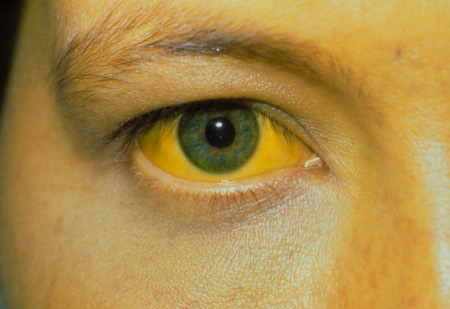
Chronic HEV infection
The physical examination in most patients with chronic HEV will be normal. Note that patients with chronic disease may progress to cirrhosis of the liver, and may develop stigmata of chronic liver disease. See Complications.
Acute liver failure/acute-on-chronic liver failure
Patients who present with acute liver failure/acute-on-chronic liver failure may have similar signs to those presenting with acute hepatitis infection; patients with acute liver failure may also have features of hepatic encephalopathy evident on examination. Hepatic encephalopathy encompasses a spectrum of neurological and psychiatric changes; initial signs and symptoms may be subtle.[55]Vilstrup H, Amodio P, Bajaj J, et al. Hepatic encephalopathy in chronic liver disease: 2014 practice guideline by the American Association for the Study of Liver Diseases and the European Association for the Study of the Liver. Hepatology. 2014 Aug;60(2):715-35.
https://aasldpubs.onlinelibrary.wiley.com/doi/10.1002/hep.27210
http://www.ncbi.nlm.nih.gov/pubmed/25042402?tool=bestpractice.com
Signs that may be present on examination due to encephalopathy include:[55]Vilstrup H, Amodio P, Bajaj J, et al. Hepatic encephalopathy in chronic liver disease: 2014 practice guideline by the American Association for the Study of Liver Diseases and the European Association for the Study of the Liver. Hepatology. 2014 Aug;60(2):715-35.
https://aasldpubs.onlinelibrary.wiley.com/doi/10.1002/hep.27210
http://www.ncbi.nlm.nih.gov/pubmed/25042402?tool=bestpractice.com
Asterixis
Hypertonia
Hyperreflexia
Clonus
Rigidity.
Patients with acute hepatic failure may also have hepatomegaly, as well as right upper quadrant tenderness. Less commonly patients may present with features of coagulopathy. See Acute liver failure.
Initial investigations
Test the patient for hepatitis E infection if they:[1]Centers for Disease Control and Prevention. Viral hepatitis: hepatitis E. Jun 2020 [internet publication].
https://www.cdc.gov/hepatitis/hev/index.htm
[2]European Association for the Study of the Liver. EASL clinical practice guidelines on hepatitis E virus infection. J Hepatol. 2018 Jun;68(6):1256-71.
https://www.journal-of-hepatology.eu/article/S0168-8278(18)30155-7/fulltext
http://www.ncbi.nlm.nih.gov/pubmed/29609832?tool=bestpractice.com
Present with an acute hepatitis and have travelled from an area in which there is a hepatitis E outbreak or where hepatitis E is endemic
Have unexplained symptoms of liver injury, regardless of travel history, and test negative for serological markers of hepatitis A, hepatitis B, hepatitis C, other hepatotropic viruses, and all other causes of acute liver injury; see Hepatitis A, Hepatitis B, Hepatitis C
Present with suspected drug-induced liver injury
Are immunosuppressed and have unexplained abnormal liver function tests
Have unexplained flares of chronic liver disease
Present with neuralgic amyotrophy or Guillain-Barré syndrome
Have abnormal liver function tests after receiving blood products (note this does not apply to those who have received blood transfusions in countries where there is routine nucleic acid testing of blood donations for HEV: for example, in the UK).
Also consider testing for HEV infection in patients with:[2]European Association for the Study of the Liver. EASL clinical practice guidelines on hepatitis E virus infection. J Hepatol. 2018 Jun;68(6):1256-71.
https://www.journal-of-hepatology.eu/article/S0168-8278(18)30155-7/fulltext
http://www.ncbi.nlm.nih.gov/pubmed/29609832?tool=bestpractice.com
Tests for HEV
Use a combination of nucleic acid amplification techniques (NAATs) and serological testing to confirm HEV infection.[1]Centers for Disease Control and Prevention. Viral hepatitis: hepatitis E. Jun 2020 [internet publication].
https://www.cdc.gov/hepatitis/hev/index.htm
[2]European Association for the Study of the Liver. EASL clinical practice guidelines on hepatitis E virus infection. J Hepatol. 2018 Jun;68(6):1256-71.
https://www.journal-of-hepatology.eu/article/S0168-8278(18)30155-7/fulltext
http://www.ncbi.nlm.nih.gov/pubmed/29609832?tool=bestpractice.com
[56]Miller JM, Binnicker MJ, Campbell S, et al; Infectious Diseases Society of America and the American Society for Microbiology. A guide to utilization of the microbiology laboratory for diagnosis of infectious diseases: 2018 update by the Infectious Diseases Society of America and the American Society for Microbiology. Clin Infect Dis. 2018 Aug 31;67(6):e1-94.
https://academic.oup.com/cid/article/67/6/e1/5046039
http://www.ncbi.nlm.nih.gov/pubmed/29955859?tool=bestpractice.com
HEV antibody (anti-HEV) and HEV RNA polymerase chain reaction (PCR) tests are the basic tests for diagnosis of HEV infection and subsequent monitoring. However, availability of these tests may be limited in some regions - follow your local protocol. In the US, for example, these tests can be ordered from the Centers for Disease Control and Prevention.
CDC: hepatitis E information - laboratory testing requests
Opens in new window Rapid tests for HEV infection detection are also available.[15]World Health Organization. Fact sheets: hepatitis E. Jul 2023 [internet publication].
https://www.who.int/en/news-room/fact-sheets/detail/hepatitis-e
Always request anti-HEV immunoglobulin M (IgM) screening in immunocompetent patients with suspected HEV.[2]European Association for the Study of the Liver. EASL clinical practice guidelines on hepatitis E virus infection. J Hepatol. 2018 Jun;68(6):1256-71.
https://www.journal-of-hepatology.eu/article/S0168-8278(18)30155-7/fulltext
http://www.ncbi.nlm.nih.gov/pubmed/29609832?tool=bestpractice.com
[15]World Health Organization. Fact sheets: hepatitis E. Jul 2023 [internet publication].
https://www.who.int/en/news-room/fact-sheets/detail/hepatitis-e
[56]Miller JM, Binnicker MJ, Campbell S, et al; Infectious Diseases Society of America and the American Society for Microbiology. A guide to utilization of the microbiology laboratory for diagnosis of infectious diseases: 2018 update by the Infectious Diseases Society of America and the American Society for Microbiology. Clin Infect Dis. 2018 Aug 31;67(6):e1-94.
https://academic.oup.com/cid/article/67/6/e1/5046039
http://www.ncbi.nlm.nih.gov/pubmed/29955859?tool=bestpractice.com
Definitive diagnosis of HEV infection in endemic areas is usually based on serum anti-HEV IgM antibody detection in immunocompetent patients.[15]World Health Organization. Fact sheets: hepatitis E. Jul 2023 [internet publication].
https://www.who.int/en/news-room/fact-sheets/detail/hepatitis-e
[56]Miller JM, Binnicker MJ, Campbell S, et al; Infectious Diseases Society of America and the American Society for Microbiology. A guide to utilization of the microbiology laboratory for diagnosis of infectious diseases: 2018 update by the Infectious Diseases Society of America and the American Society for Microbiology. Clin Infect Dis. 2018 Aug 31;67(6):e1-94.
https://academic.oup.com/cid/article/67/6/e1/5046039
http://www.ncbi.nlm.nih.gov/pubmed/29955859?tool=bestpractice.com
Anti-HEV IgM is usually detectable from 1 week to 2 months after exposure to the virus and typically persists for 3-4 months.[2]European Association for the Study of the Liver. EASL clinical practice guidelines on hepatitis E virus infection. J Hepatol. 2018 Jun;68(6):1256-71.
https://www.journal-of-hepatology.eu/article/S0168-8278(18)30155-7/fulltext
http://www.ncbi.nlm.nih.gov/pubmed/29609832?tool=bestpractice.com
[56]Miller JM, Binnicker MJ, Campbell S, et al; Infectious Diseases Society of America and the American Society for Microbiology. A guide to utilization of the microbiology laboratory for diagnosis of infectious diseases: 2018 update by the Infectious Diseases Society of America and the American Society for Microbiology. Clin Infect Dis. 2018 Aug 31;67(6):e1-94.
https://academic.oup.com/cid/article/67/6/e1/5046039
http://www.ncbi.nlm.nih.gov/pubmed/29955859?tool=bestpractice.com
Some organisations, such as the European Association for the Study of the Liver (EASL), also recommend anti-HEV IgG screening alongside anti-HEV IgM.[2]European Association for the Study of the Liver. EASL clinical practice guidelines on hepatitis E virus infection. J Hepatol. 2018 Jun;68(6):1256-71.
https://www.journal-of-hepatology.eu/article/S0168-8278(18)30155-7/fulltext
http://www.ncbi.nlm.nih.gov/pubmed/29609832?tool=bestpractice.com
Bear in mind, however, that anti-HEV IgG positivity may reflect past infection. Anti-HEV IgG is detectable later in the clinical course than anti-HEV IgM; the titre increases throughout the illness, and can persist for many years.[2]European Association for the Study of the Liver. EASL clinical practice guidelines on hepatitis E virus infection. J Hepatol. 2018 Jun;68(6):1256-71.
https://www.journal-of-hepatology.eu/article/S0168-8278(18)30155-7/fulltext
http://www.ncbi.nlm.nih.gov/pubmed/29609832?tool=bestpractice.com
If the patient is immunosuppressed, use other tests, such as reverse transcriptase polymerase chain reaction (RT-PCR) for hepatitis E virus RNA detection in serum and stool (note these tests require specialised testing facilities).[2]European Association for the Study of the Liver. EASL clinical practice guidelines on hepatitis E virus infection. J Hepatol. 2018 Jun;68(6):1256-71.
https://www.journal-of-hepatology.eu/article/S0168-8278(18)30155-7/fulltext
http://www.ncbi.nlm.nih.gov/pubmed/29609832?tool=bestpractice.com
[15]World Health Organization. Fact sheets: hepatitis E. Jul 2023 [internet publication].
https://www.who.int/en/news-room/fact-sheets/detail/hepatitis-e
[25]British Transplantation Society. Guidelines for hepatitis E and solid organ transplantation. First edition. Apr 2017 [internet publication].
https://bts.org.uk/wp-content/uploads/2017/06/BTS_HEV_Guideline-FINAL.pdf
[41]Te H, Doucette K. Viral hepatitis: guidelines by the American Society of Transplantation Infectious Disease Community of Practice. Clin Transplant. 2019 Sep;33(9):e13514.
http://www.ncbi.nlm.nih.gov/pubmed/30817047?tool=bestpractice.com
[56]Miller JM, Binnicker MJ, Campbell S, et al; Infectious Diseases Society of America and the American Society for Microbiology. A guide to utilization of the microbiology laboratory for diagnosis of infectious diseases: 2018 update by the Infectious Diseases Society of America and the American Society for Microbiology. Clin Infect Dis. 2018 Aug 31;67(6):e1-94.
https://academic.oup.com/cid/article/67/6/e1/5046039
http://www.ncbi.nlm.nih.gov/pubmed/29955859?tool=bestpractice.com
Antibody detection is unreliable in transplant recipients and other people who are immunosuppressed, due to delayed or impaired humoral response (which may lead to false negative results).[2]European Association for the Study of the Liver. EASL clinical practice guidelines on hepatitis E virus infection. J Hepatol. 2018 Jun;68(6):1256-71.
https://www.journal-of-hepatology.eu/article/S0168-8278(18)30155-7/fulltext
http://www.ncbi.nlm.nih.gov/pubmed/29609832?tool=bestpractice.com
[25]British Transplantation Society. Guidelines for hepatitis E and solid organ transplantation. First edition. Apr 2017 [internet publication].
https://bts.org.uk/wp-content/uploads/2017/06/BTS_HEV_Guideline-FINAL.pdf
[41]Te H, Doucette K. Viral hepatitis: guidelines by the American Society of Transplantation Infectious Disease Community of Practice. Clin Transplant. 2019 Sep;33(9):e13514.
http://www.ncbi.nlm.nih.gov/pubmed/30817047?tool=bestpractice.com
[56]Miller JM, Binnicker MJ, Campbell S, et al; Infectious Diseases Society of America and the American Society for Microbiology. A guide to utilization of the microbiology laboratory for diagnosis of infectious diseases: 2018 update by the Infectious Diseases Society of America and the American Society for Microbiology. Clin Infect Dis. 2018 Aug 31;67(6):e1-94.
https://academic.oup.com/cid/article/67/6/e1/5046039
http://www.ncbi.nlm.nih.gov/pubmed/29955859?tool=bestpractice.com
[57]Kamar N, Rostaing L, Abravanel F, et al. Ribavirin therapy inhibits viral replication on patients with chronic hepatitis E virus infection. Gastroenterology. 2010 Nov;139(5):1612-8.
https://www.gastrojournal.org/article/S0016-5085(10)01169-8/fulltext
http://www.ncbi.nlm.nih.gov/pubmed/20708006?tool=bestpractice.com
These approaches may also be helpful for diagnosis in areas where HEV infection is less common, and to detect chronic HEV infection.[15]World Health Organization. Fact sheets: hepatitis E. Jul 2023 [internet publication].
https://www.who.int/en/news-room/fact-sheets/detail/hepatitis-e
HEV RNA present in serum or stool indicates HEV infection and is often the only positive test in immunosuppressed patients with chronic HEV infection.[56]Miller JM, Binnicker MJ, Campbell S, et al; Infectious Diseases Society of America and the American Society for Microbiology. A guide to utilization of the microbiology laboratory for diagnosis of infectious diseases: 2018 update by the Infectious Diseases Society of America and the American Society for Microbiology. Clin Infect Dis. 2018 Aug 31;67(6):e1-94.
https://academic.oup.com/cid/article/67/6/e1/5046039
http://www.ncbi.nlm.nih.gov/pubmed/29955859?tool=bestpractice.com
HEV RNA is detectable around 3 weeks after onset of infection, and may persist for several weeks after infection has cleared.[2]European Association for the Study of the Liver. EASL clinical practice guidelines on hepatitis E virus infection. J Hepatol. 2018 Jun;68(6):1256-71.
https://www.journal-of-hepatology.eu/article/S0168-8278(18)30155-7/fulltext
http://www.ncbi.nlm.nih.gov/pubmed/29609832?tool=bestpractice.com
[17]Kamar N, Bendall R, Legrand-Abravanel F, et al. Hepatitis E. Lancet. 2012 Jun 30;379(9835):2477-88.
http://www.ncbi.nlm.nih.gov/pubmed/22549046?tool=bestpractice.com
Chronic HEV infection is defined by the persistence of HEV RNA for 3 or more months.[2]European Association for the Study of the Liver. EASL clinical practice guidelines on hepatitis E virus infection. J Hepatol. 2018 Jun;68(6):1256-71.
https://www.journal-of-hepatology.eu/article/S0168-8278(18)30155-7/fulltext
http://www.ncbi.nlm.nih.gov/pubmed/29609832?tool=bestpractice.com
[4]Kamar N, Selves J, Mansuy JM, et al. Hepatitis E virus and chronic hepatitis in organ-transplant recipients. N Engl J Med. 2008 Feb 21;358(8):811-7.
https://www.nejm.org/doi/full/10.1056/NEJMoa0706992
http://www.ncbi.nlm.nih.gov/pubmed/18287603?tool=bestpractice.com
[5]Marion O, Abravanel F, Lhomme S, et al. Hepatitis E in transplantation. Curr Infect Dis Rep. 2016 Mar;18(3):8.
http://www.ncbi.nlm.nih.gov/pubmed/26838163?tool=bestpractice.com
In practice, serum HEV RNA testing is often used in preference to stool analysis due to availability and ease of testing; however, stool testing may be useful following treatment of chronic infection as a negative stool HEV RNA is helpful to confirm treatment success.
Characterisation of liver status
Perform the following laboratory tests in all patients to characterise liver disease status:
Consider abdominal ultrasound as a first imaging test in patients who may have underlying liver disease, are immunocompromised, or have chronic infection to evaluate the liver for the presence of fibrosis, cirrhosis, and portal hypertension, and to exclude hepatocellular carcinoma.
Other investigations
Triphasic contrast computed tomography or contrast magnetic resonance imaging may be considered, particularly in patients with advanced fibrosis or cirrhosis. Non-invasive methods of assessing liver fibrosis (e.g., transient elastography using magnetic resonance or Fibroscan, fibrosis biomarkers) or liver biopsy can be obtained in situations where there is a diagnostic dilemma, but should not be routinely required.
Consider specialist nephrology review for further renal investigation if the patient has recent deterioration of renal function or significant proteinuria.[2]European Association for the Study of the Liver. EASL clinical practice guidelines on hepatitis E virus infection. J Hepatol. 2018 Jun;68(6):1256-71.
https://www.journal-of-hepatology.eu/article/S0168-8278(18)30155-7/fulltext
http://www.ncbi.nlm.nih.gov/pubmed/29609832?tool=bestpractice.com
Renal manifestations of HEV infection include pre-renal failure, glomerular disorders, and tubular and interstitial injury.[58]Kovvuru K, Carbajal N, Pakanati AR, et al. Renal manifestations of hepatitis E among immunocompetent and solid organ transplant recipients. World J Hepatol. 2022 Mar 27;14(3):516-24.
https://www.wjgnet.com/1948-5182/full/v14/i3/516.htm
http://www.ncbi.nlm.nih.gov/pubmed/35582296?tool=bestpractice.com
Consider referral to a neurologist for further investigations if the patient presents with neurological manifestations related to HEV infection.