The vast majority of clavicle fractures result from acute trauma due to a direct fall/impact onto the shoulder (e.g., while playing sports), a motor vehicle accident, or another high-energy trauma (e.g., fall from a height). Much less commonly, patients present following a fall on the outstretched hand or a direct blow to the clavicle itself.
Patients are generally young males (<30 years), although older women (≥65 years) are at higher risk for low-energy mechanism clavicle fractures.[5]Kihlström C, Möller M, Lönn K, et al. Clavicle fractures: epidemiology, classification and treatment of 2 422 fractures in the Swedish Fracture Register; an observational study. BMC Musculoskelet Disord. 2017 Feb 15;18(1):82.
https://www.doi.org/10.1186/s12891-017-1444-1
http://www.ncbi.nlm.nih.gov/pubmed/28202071?tool=bestpractice.com
History
Patients presenting with an acute clavicle fracture typically report moderate to severe pain at the fracture site. The pain is worsened by shoulder range of motion or trying to use the injured extremity.
The patient may describe an audible crack at the time of injury and/or palpable crepitus, especially during attempted range of motion of the shoulder. They may report swelling and/or obvious deformity in the region of the injury.
Ask about the mechanism of injury. Almost all clavicle fractures are caused by acute trauma; stress fractures of the clavicle are rare.[20]Miyamoto S, Otsuka M, Hasue F, et al. Stress fracture of the midshaft clavicle associated with sternocostoclavicular hyperostosis-Case report. Int J Surg Case Rep. 2019;58:121-6.
https://www.doi.org/10.1016/j.ijscr.2019.03.059
http://www.ncbi.nlm.nih.gov/pubmed/31035227?tool=bestpractice.com
The most common mechanisms of injury include:
Direct fall on the shoulder (e.g., while playing sports)[5]Kihlström C, Möller M, Lönn K, et al. Clavicle fractures: epidemiology, classification and treatment of 2 422 fractures in the Swedish Fracture Register; an observational study. BMC Musculoskelet Disord. 2017 Feb 15;18(1):82.
https://www.doi.org/10.1186/s12891-017-1444-1
http://www.ncbi.nlm.nih.gov/pubmed/28202071?tool=bestpractice.com
[6]Song HS, Kim H. Current concepts in the treatment of midshaft clavicle fractures in adults. Clin Shoulder Elb. 2021 Sep;24(3):189-98.
https://www.doi.org/10.5397/cise.2021.00388
http://www.ncbi.nlm.nih.gov/pubmed/34488301?tool=bestpractice.com
[14]Stanley D, Norris SH. Recovery following fractures of the clavicle treated conservatively. Injury. 1988 May;19(3):162-4.
http://www.ncbi.nlm.nih.gov/pubmed/3248891?tool=bestpractice.com
[15]Pecci M, Kreher JB. Clavicle fractures. Am Fam Physician. 2008 Jan 1;77(1):65-70.
http://www.ncbi.nlm.nih.gov/pubmed/18236824?tool=bestpractice.com
[16]Nowak J, Mallmin H, Larsson S. The aetiology and epidemiology of clavicular fractures. A prospective study during a two-year period in Uppsala, Sweden. Injury. 2000 Jun;31(5):353-8.
http://www.ncbi.nlm.nih.gov/pubmed/10775691?tool=bestpractice.com
Motor vehicle accidents (or other high-energy trauma)[3]Robinson CM. Fractures of the clavicle in the adult. Epidemiology and classification. J Bone Joint Surg Br. 1998 May;80(3):476-84.
https://www.doi.org/10.1302/0301-620x.80b3.8079
http://www.ncbi.nlm.nih.gov/pubmed/9619941?tool=bestpractice.com
[5]Kihlström C, Möller M, Lönn K, et al. Clavicle fractures: epidemiology, classification and treatment of 2 422 fractures in the Swedish Fracture Register; an observational study. BMC Musculoskelet Disord. 2017 Feb 15;18(1):82.
https://www.doi.org/10.1186/s12891-017-1444-1
http://www.ncbi.nlm.nih.gov/pubmed/28202071?tool=bestpractice.com
[6]Song HS, Kim H. Current concepts in the treatment of midshaft clavicle fractures in adults. Clin Shoulder Elb. 2021 Sep;24(3):189-98.
https://www.doi.org/10.5397/cise.2021.00388
http://www.ncbi.nlm.nih.gov/pubmed/34488301?tool=bestpractice.com
Fall from a bicycle or horse[6]Song HS, Kim H. Current concepts in the treatment of midshaft clavicle fractures in adults. Clin Shoulder Elb. 2021 Sep;24(3):189-98.
https://www.doi.org/10.5397/cise.2021.00388
http://www.ncbi.nlm.nih.gov/pubmed/34488301?tool=bestpractice.com
[16]Nowak J, Mallmin H, Larsson S. The aetiology and epidemiology of clavicular fractures. A prospective study during a two-year period in Uppsala, Sweden. Injury. 2000 Jun;31(5):353-8.
http://www.ncbi.nlm.nih.gov/pubmed/10775691?tool=bestpractice.com
Direct trauma to the clavicle (e.g., being struck by a hard object, such as a baseball bat)[6]Song HS, Kim H. Current concepts in the treatment of midshaft clavicle fractures in adults. Clin Shoulder Elb. 2021 Sep;24(3):189-98.
https://www.doi.org/10.5397/cise.2021.00388
http://www.ncbi.nlm.nih.gov/pubmed/34488301?tool=bestpractice.com
Fall on the outstretched hand.[14]Stanley D, Norris SH. Recovery following fractures of the clavicle treated conservatively. Injury. 1988 May;19(3):162-4.
http://www.ncbi.nlm.nih.gov/pubmed/3248891?tool=bestpractice.com
[15]Pecci M, Kreher JB. Clavicle fractures. Am Fam Physician. 2008 Jan 1;77(1):65-70.
http://www.ncbi.nlm.nih.gov/pubmed/18236824?tool=bestpractice.com
Other risk factors to consider include:
Male sex: males are more than twice as likely as females to sustain a clavicle fracture overall.[5]Kihlström C, Möller M, Lönn K, et al. Clavicle fractures: epidemiology, classification and treatment of 2 422 fractures in the Swedish Fracture Register; an observational study. BMC Musculoskelet Disord. 2017 Feb 15;18(1):82.
https://www.doi.org/10.1186/s12891-017-1444-1
http://www.ncbi.nlm.nih.gov/pubmed/28202071?tool=bestpractice.com
[6]Song HS, Kim H. Current concepts in the treatment of midshaft clavicle fractures in adults. Clin Shoulder Elb. 2021 Sep;24(3):189-98.
https://www.doi.org/10.5397/cise.2021.00388
http://www.ncbi.nlm.nih.gov/pubmed/34488301?tool=bestpractice.com
Age 15-24 years: clavicle fractures in this age group are often associated with a high-energy mechanism or sports injury.[5]Kihlström C, Möller M, Lönn K, et al. Clavicle fractures: epidemiology, classification and treatment of 2 422 fractures in the Swedish Fracture Register; an observational study. BMC Musculoskelet Disord. 2017 Feb 15;18(1):82.
https://www.doi.org/10.1186/s12891-017-1444-1
http://www.ncbi.nlm.nih.gov/pubmed/28202071?tool=bestpractice.com
[15]Pecci M, Kreher JB. Clavicle fractures. Am Fam Physician. 2008 Jan 1;77(1):65-70.
http://www.ncbi.nlm.nih.gov/pubmed/18236824?tool=bestpractice.com
Age ≥65 years in females: older age is a risk factor for low-energy mechanism clavicle fractures. (i.e., ground-level fall).[5]Kihlström C, Möller M, Lönn K, et al. Clavicle fractures: epidemiology, classification and treatment of 2 422 fractures in the Swedish Fracture Register; an observational study. BMC Musculoskelet Disord. 2017 Feb 15;18(1):82.
https://www.doi.org/10.1186/s12891-017-1444-1
http://www.ncbi.nlm.nih.gov/pubmed/28202071?tool=bestpractice.com
Patients with osteoporosis, other metabolic bone disease, or various malignancies are at increased risk for pathological fracture, which may occur with what appears to be minimal trauma or trivial provocation.[18]Priemel MH, Stiel N, Zustin J, et al. Bone tumours of the clavicle: histopathological, anatomical and epidemiological analysis of 113 cases. J Bone Oncol. 2019 Jun;16:100229.
https://www.doi.org/10.1016/j.jbo.2019.100229
http://www.ncbi.nlm.nih.gov/pubmed/30976505?tool=bestpractice.com
Low body mass index, a previous fracture, and corticosteroid use are weak risk factors for an insufficiency fracture in general, though not specifically a clavicle fracture.
Neoplasm, infection, and arthritis may mimic a clavicle fracture.[18]Priemel MH, Stiel N, Zustin J, et al. Bone tumours of the clavicle: histopathological, anatomical and epidemiological analysis of 113 cases. J Bone Oncol. 2019 Jun;16:100229.
https://www.doi.org/10.1016/j.jbo.2019.100229
http://www.ncbi.nlm.nih.gov/pubmed/30976505?tool=bestpractice.com
See Differentials.
Physical examination
Complete a trauma survey, especially in the event of high-energy trauma (e.g., motor vehicle accident). Assess for underlying and associated serious injuries with an ABCDE primary trauma survey:[21]Committee on Trauma, American College of Surgeons. ATLS: advanced trauma life support program for doctors. 8th ed. Chicago, IL: American College of Surgeons; 2008.
Airway with cervical spine precautions
Breathing and ventilation
Circulation with haemorrhage control
Disability (assess neurological status)
Exposure (ensure no injury is missed) and Environmental control.
Signs of vascular injury include a diminished pulse (compared with the contralateral uninjured side), pallor, and/or cool or cold tissues in the affected extremity. Hypotension, tachycardia out of proportion to pain, or a decreased or decreasing level of consciousness imply severe blood loss and shock.
However, these signs could also be due to other injuries, such as associated tension pneumothorax or intracranial injury, underlining the great importance of a thorough trauma survey and careful evaluation.
Focused examination of clavicle and shoulder girdle
Once the trauma survey has been completed and the patient is determined to be clinically stable without more serious or urgent associated injury, perform a focused examination of the clavicle and shoulder girdle.
Inspection usually reveals soft tissue swelling around the clavicle and ecchymosis is often present within a day or two after the injury, gradually diminishing over time. An obvious deformity may be present if the fracture is significantly displaced and/or angulated, as often happens in a mid-clavicle fracture due to the deforming forces on the bones from the associated musculature and weight of the arm. The sternocleidomastoid exerts upward force on the proximal fragment and the trapezius pulls the distal fragment upwards, whereas the clavicular head of the pectoralis major (along with indirect pull from the latissimus dorsi) exerts downward and medial force on the distal fragment. Combined with the downward directed force from the weight of the arm, these forces often result in displacement of the fracture with the proximal end being pulled superiorly, the distal end being pulled inferiorly, and shortening as the distal fragment is pulled inferomedially.[15]Pecci M, Kreher JB. Clavicle fractures. Am Fam Physician. 2008 Jan 1;77(1):65-70.
http://www.ncbi.nlm.nih.gov/pubmed/18236824?tool=bestpractice.com
Look carefully for skin tenting. Although this is uncommon (with studies showing an incidence of 4% to 9% in patients with displaced midshaft clavicle fracture), skin tenting is a risk factor for skin necrosis and may lead to an open injury.[22]Chalmers PN, Van Thiel GS, Ferry ST. Is skin tenting secondary to displaced clavicle fracture more than a theoretical risk? A report of 2 adolescent cases. Am J Orthop (Belle Mead NJ). 2015 Oct;44(10):E414-6.
http://www.ncbi.nlm.nih.gov/pubmed/26447424?tool=bestpractice.com
[23]Kirmani SJ, Pillai SK, Madegowda BR, et al. Vertical fragment in adult midshaft clavicle fractures: an indicator for surgical intervention. Orthopedics. 2009 Oct;32(10).
http://www.ncbi.nlm.nih.gov/pubmed/19824609?tool=bestpractice.com
[24]Zhang D, Earp BE, Dyer GSM. Skin tenting in displaced midshaft clavicle fractures. Arch Bone Jt Surg. 2021 Jul;9(4):418-22.
http://www.ncbi.nlm.nih.gov/pubmed/34423090?tool=bestpractice.com
It therefore requires urgent orthopaedic consultation.[25]Monica J, Vredenburgh Z, Korsh J, et al. Acute shoulder injuries in adults. Am Fam Physician. 2016 Jul 15;94(2):119-27.
http://www.ncbi.nlm.nih.gov/pubmed/27419328?tool=bestpractice.com
If a wound is noted at or near the fracture site, treat the injury as an open fracture and refer for immediate orthopaedic consultation.
Look for impaired movement of the shoulder and guarding against movement of the upper limb. Patients tend to keep the injured arm at their side, and often try to support the weight of the injured extremity with their contralateral hand or by resting it on a table or the arm of a chair.
Stress fractures also present with pain on motion and tenderness to palpation.
Assess the patient’s pain regularly and consider this during the physical examination. Palpate gently over the suspected site of injury. This will usually elicit point tenderness at the fracture site, often with associated crepitus. Carefully palpate over the entire clavicle, including to the acromioclavicular and sternoclavicular joints. The vast majority (69% to 82%) of clavicle fractures involve the mid-clavicle, and up to 90% of these fractures have some degree of displacement or angulation.[5]Kihlström C, Möller M, Lönn K, et al. Clavicle fractures: epidemiology, classification and treatment of 2 422 fractures in the Swedish Fracture Register; an observational study. BMC Musculoskelet Disord. 2017 Feb 15;18(1):82.
https://www.doi.org/10.1186/s12891-017-1444-1
http://www.ncbi.nlm.nih.gov/pubmed/28202071?tool=bestpractice.com
Carefully assess for evidence of acromioclavicular joint instability or sternoclavicular joint instability. Although sternoclavicular joint instability is rare and tends to occur only with high-energy trauma, posterior sternoclavicular joint instability and/or a posteriorly displaced medial clavicle fracture can compromise the great vessels, nerves, and airway, and is a sign of possible additional thoracic injury, so it must not be missed.[26]Frias M, Ramos R, Bernardes M, et al. Medial clavicle fracture dislocation surgically treated: case report. Trauma Cases Rev. 2018;4:066.
https://clinmedjournals.org/articles/tcr/trauma-cases-and-reviews-tcr-4-066.php?jid=tcr
Thorough palpation of the scapula, ribcage, and cervical and thoracic spine is also necessary, as injury to these areas is not uncommon, especially in high-energy injuries. See Acute cervical spine trauma.
Diligently conducting a comprehensive physical examination of skin integrity and all pertinent neurovascular structures within the zone of injury is the best practice for the assessment of shoulder injuries. A systematic, thorough physical examination will allow the clinician to appropriately evaluate clavicular fractures and shoulder trauma patients without missing any important pathology. Assess for brachial plexus injury or damage to subclavian vessels, and auscultate the chest to assess for underlying lung injury.[15]Pecci M, Kreher JB. Clavicle fractures. Am Fam Physician. 2008 Jan 1;77(1):65-70.
http://www.ncbi.nlm.nih.gov/pubmed/18236824?tool=bestpractice.com
Perform a neurovascular examination of the affected extremity and consider referral as needed. Check for any other injuries.[25]Monica J, Vredenburgh Z, Korsh J, et al. Acute shoulder injuries in adults. Am Fam Physician. 2016 Jul 15;94(2):119-27.
http://www.ncbi.nlm.nih.gov/pubmed/27419328?tool=bestpractice.com
Associated conditions not to miss
In all cases of clavicle fracture, the physician should evaluate for other rare but potentially dangerous concomitant injuries, such as sternoclavicular dislocation or scapulothoracic dissociation.
Anterior and posterior sternoclavicular dislocations are rare injuries resulting from high-energy trauma.[27]Tompkins M, Bliss J, Villarreal R, et al. Posterior sternoclavicular disruption with ipsilateral clavicle fracture in a nine-year-old hockey player. J Orthop Trauma. 2010 Apr;24(4):e36-9.
http://www.ncbi.nlm.nih.gov/pubmed/20335749?tool=bestpractice.com
They are generally associated with thoracic injury. A posterior dislocation of the medial clavicle can be fatal in that it can injure adjacent neurovascular structures, the oesophagus and/or trachea as well as cause pneumothorax or haemothorax injuries.[28]O'Connor PA, Nölke L, O'Donnell A, et al. Retrosternal dislocation of the clavicle associated with a traumatic pneumothorax. Interact Cardiovasc Thorac Surg. 2003 Mar;2(1):9-11.
https://www.doi.org/10.1016/S1569-9293(02)00066-X
http://www.ncbi.nlm.nih.gov/pubmed/17669976?tool=bestpractice.com
[29]Yadav S, Khanna V, Mukherjee S. Ipsilateral sternoclavicular joint anterior dislocation with fracture of the mid shaft of the clavicle. J Clin Orthop Trauma. 2019 May-Jun;10(3):510-3.
https://www.doi.org/10.1016/j.jcot.2018.08.002
http://www.ncbi.nlm.nih.gov/pubmed/31061579?tool=bestpractice.com
The patient may present with dysphagia, stridor, asymmetric pulses, or paresthesia due to compression of mediastinal structures. Concern for this injury requires an urgent CT scan of the area to assess the status of the medial clavicle, sternoclavicular joint, and associated neurovascular structures and soft tissues.
Scapulothoracic dissociation is another rare entity that can be associated with a displaced clavicle fracture. It consists of disruption of the scapulothoracic articulation, usually due to high-energy trauma with lateral traction to the shoulder girdle. Scapulothoracic dissociation can be associated with bony injury such as a displaced clavicle fracture as well as severe neurovascular injury to the brachial plexus, the axillary artery, or the subclavian artery.[30]Ebraheim NA, An HS, Jackson WT, et al. Scapulothoracic dissociation. J Bone Joint Surg Am. 1988 Mar;70(3):428-32.
http://www.ncbi.nlm.nih.gov/pubmed/3346268?tool=bestpractice.com
[31]Clements RH, Reisser JR. Scapulothoracic dissociation: a devastating injury. J Trauma. 1996 Jan;40(1):146-9.
http://www.ncbi.nlm.nih.gov/pubmed/8576982?tool=bestpractice.com
Initial investigations
Clavicle x-ray series
Order clavicle x-rays for all patients with suspected clavicle fracture.[25]Monica J, Vredenburgh Z, Korsh J, et al. Acute shoulder injuries in adults. Am Fam Physician. 2016 Jul 15;94(2):119-27.
http://www.ncbi.nlm.nih.gov/pubmed/27419328?tool=bestpractice.com
[32]American College of Radiology. ACR appropriateness criteria: acute shoulder pain. 2024 [internet publication].
https://acsearch.acr.org/docs/69433/narrative
A standard clavicle x-ray series includes anteroposterior (AP) views in internal and external rotation and an axillary or scapula-Y view.[32]American College of Radiology. ACR appropriateness criteria: acute shoulder pain. 2024 [internet publication].
https://acsearch.acr.org/docs/69433/narrative
Axillary or scapula-Y views are vital in evaluating traumatic shoulder injuries as acromioclavicular and glenohumeral joint dislocations can be misclassified on AP views.[32]American College of Radiology. ACR appropriateness criteria: acute shoulder pain. 2024 [internet publication].
https://acsearch.acr.org/docs/69433/narrative
[33]Vaisman A, Villalón Montenegro IE, Tuca De Diego MJ, et al. A novel radiographic index for the diagnosis of posterior acromioclavicular joint dislocations. Am J Sports Med. 2014 Jan;42(1):112-6.
http://www.ncbi.nlm.nih.gov/pubmed/24124199?tool=bestpractice.com
[34]Emond M, Le Sage N, Lavoie A, et al. Refinement of the Quebec decision rule for radiography in shoulder dislocation. CJEM. 2009 Jan;11(1):36-43.
http://www.ncbi.nlm.nih.gov/pubmed/19166638?tool=bestpractice.com
The exact angle used for the cephalic tilt view depends on patient positioning and body habitus to ensure the clavicle is projected clearly above the scapula and 2nd and 3rd ribs. Position the patient upright, rather than supine, as this may better demonstrate the degree of any displacement in a midshaft fracture.[35]American Academy of Orthopaedic Surgeons. Treatment of clavicle fractures. Evidence-based clinical practice guideline. Dec 2022 [internet publication].
https://www.aaos.org/globalassets/quality-and-practice-resources/clavicle-fractures/clavicle-fractures-cpg.pdf
[Figure caption and citation for the preceding image starts]: Anteroposterior radiograph of right shoulder demonstrating clavicle fractureArnold S et al. BMJ Case Reports CP 2021;14:e241382; used with permission [Citation ends].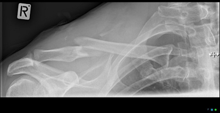
[Figure caption and citation for the preceding image starts]: Anteroposterior radiograph of left shoulder demonstrating a clavicle fractureAlao D et al. Emergency Medicine Journal 2005;22:232-3; used with permission [Citation ends].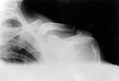
Computed tomography (CT) of chest, abdomen, pelvis
A chest CT may be required as an initial investigation in patients where there is a high degree of suspicion of a thoracic injury, such as in a polytrauma patient.[36]Wright J, Aresti N, Heuveling C, et al. Are standard antero-posterior and 20° caudal radiographs a true assessment of mid-shaft clavicular fracture displacement? J Clin Orthop Trauma. 2016 Oct-Dec;7(4):221-4.
https://www.doi.org/10.1016/j.jcot.2015.06.004
http://www.ncbi.nlm.nih.gov/pubmed/27857493?tool=bestpractice.com
Consider any associated injuries, which would lower the threshold for a trauma CT series of chest, abdomen, and pelvis.[37]Amer KM, Congiusta DV, Suri P, et al. Clavicle fractures: associated trauma and morbidity. J Clin Orthop Trauma. 2021 Feb;13:53-6.
https://www.doi.org/10.1016/j.jcot.2020.08.020
http://www.ncbi.nlm.nih.gov/pubmed/33717875?tool=bestpractice.com
Other investigations
X-rays at other sites
Order a chest x-ray posteroanterior view if there is suspected shortening of the clavicle fracture (to enable comparison with the opposite side) and/or suspected associated rib fracture.
Order a scapula x-ray series if there is a suspected scapula fracture.
Order a shoulder x-ray series (anteroposterior internal and external rotation, scapular 'Y', and axillary lateral views if there is suspected injury to the proximal humerus or glenohumeral joint).[32]American College of Radiology. ACR appropriateness criteria: acute shoulder pain. 2024 [internet publication].
https://acsearch.acr.org/docs/69433/narrative
This will reveal a humeral fracture and dislocation of the glenohumeral joint.
Clavicle CT
CT is not usually needed for most clavicle fractures and generally should be reserved for cases in which the clinician suspects a more complex injury than the plain radiographs show (such as a sternoclavicular joint dislocation).[32]American College of Radiology. ACR appropriateness criteria: acute shoulder pain. 2024 [internet publication].
https://acsearch.acr.org/docs/69433/narrative
[38]Morell DJ, Thyagarajan DS. Sternoclavicular joint dislocation and its management: A review of the literature. World J Orthop. 2016 Apr 18;7(4):244-50.
https://www.doi.org/10.5312/wjo.v7.i4.244
http://www.ncbi.nlm.nih.gov/pubmed/27114931?tool=bestpractice.com
A CT will give an accurate estimate of shortening and displacement, and may impact treatment planning.
Ultrasound
Ultrasound is rarely indicated in the evaluation of clavicle fractures.[32]American College of Radiology. ACR appropriateness criteria: acute shoulder pain. 2024 [internet publication].
https://acsearch.acr.org/docs/69433/narrative
It may be useful for detection of fracture-associated haematoma. It can show fracture at the surface of bone if present and/or joint instability if there is a high index of suspicion, but the injuries are not seen on plain x-rays. It may be useful if trying to avoid radiation exposure in a patient, such as in a child or a pregnant patient.[39]Halm BM, Chaudoin LT. Diagnosis of a posterior fracture dislocation of the medial clavicle in an adolescent with point-of-care ultrasound. Pediatr Emerg Care. 2017 Jul;33(7):519-21.
http://www.ncbi.nlm.nih.gov/pubmed/28419018?tool=bestpractice.com
Magnetic resonance imaging
Magnetic resonance imaging is usually not indicated but may help with surgical planning for high-grade injuries.[40]Flores DV, Goes PK, Gómez CM, et al. Imaging of the acromioclavicular joint: anatomy, function, pathologic features, and treatment. Radiographics. 2020 Sep-Oct;40(5):1355-82.
https://www.doi.org/10.1148/rg.2020200039
http://www.ncbi.nlm.nih.gov/pubmed/32762593?tool=bestpractice.com
It also may help to more accurately prognosticate return to activity in athletes with sprains at the acromioclavicular joint.[41]White LM, Ehmann J, Bleakney RR, et al. Acromioclavicular joint injuries in professional ice hockey players: epidemiologic and MRI findings and association with return to play. Orthop J Sports Med. 2020 Nov;8(11):2325967120964474.
https://www.doi.org/10.1177/2325967120964474
http://www.ncbi.nlm.nih.gov/pubmed/33283007?tool=bestpractice.com