Images and videos
Images


Xeroderma pigmentosum
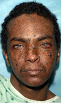
A 23 year old man from northern Africa with XP (subtype XPC) presenting with numerous hyperpigmented macules on his face. Nodular basal cell cancer is present on his left nasal root. Pigmented basal cell cancer is present on his left cheek. His eyes show corneal scarring from unprotected sun exposure
Bradford PT et al. J Med Genet 2011;48:168-76; used with permission
See this image in context in the following section/s:

Xeroderma pigmentosum
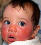
A child with XP (subtype XPD) at 9 months of age with severe blistering erythema of the malar area following minimal sun exposure. Note the sparing of her forehead and eyes that were protected by a hat
Bradford PT et al. J Med Genet 2011;48:168-76; used with permission
See this image in context in the following section/s:
Xeroderma pigmentosum
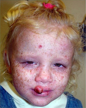
A child with XP (subtype XPC) at age 2 years. Her parents reported that she did not sunburn easily, but she developed multiple hyperpigmented macules on her face. A rapidly growing squamous cell carcinoma or keratoacanthoma grew on her upper lip and a pre-cancerous lesion appeared on her forehead
Bradford PT et al. J Med Genet 2011;48:168-76; used with permission
See this image in context in the following section/s:

Xeroderma pigmentosum
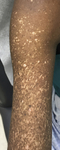
Poikilodermatous changes of the upper extremity in an 18 year old woman presenting with XP. The changes had been present since early childhood with progressive increase in size and number. A diagnosis of freckles had initially been made
Plante J et al. JAAD Case Rep 2021;13:141-43; used with permission
See this image in context in the following section/s:
Xeroderma pigmentosum
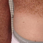
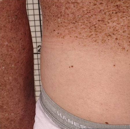
Relatively protected skin on buttocks (covered by double clothing layers) of 35-year old man shows sparing of the pigmented skin lesions (lentigos) of photodamage
Rizza ERH et al. J Invest Dermatol 2021 Apr;141(4S):976-84; used with permission
See this image in context in the following section/s:
Use of this content is subject to our disclaimer