Recommendations
Urgent
Urgently refer or admit to hospital anyone with suspected acute cholecystitis.[27]
Think 'Could this be sepsis?' based on acute deterioration in an adult patient in whom there is clinical evidence or strong suspicion of infection.[28][29][30] See Sepsis in adults.
Use a systematic approach (e.g., National Early Warning Score 2 [NEWS2]), alongside your clinical judgement, for assessment; urgently consult a senior clinical decision-maker (e.g., ST4 level doctor in the UK) if you suspect sepsis.[29][30][31][32]
Refer to local guidelines for the recommended approach at your institution for assessment and management of the patient with suspected sepsis.
Having enacted a sepsis management bundle in line with the recommended approach at your institution, identify the cause. Sepsis, organ failure, or death may be caused by other illnesses or complications of cholecystitis, such as:
Acute pancreatitis
Perforated peptic ulcer
Emphysematous cholecystitis
Gangrenous cholecystitis
Gallbladder perforation.
Measure serum lipase/amylase to exclude acute pancreatitis.[27][33]
Source control is essential in patients with sepsis. Involve the surgical team early.[30] Consider immediate surgical treatment for emphysematous or gangrenous cholecystitis. Empyema may need percutaneous drainage.[3]
Confirm the diagnosis of acute cholecystitis using ultrasound.[3][27][34]
Key Recommendations
Presentation
Patients typically present with pain and localised tenderness, with or without guarding, in the upper right quadrant.
There may be evidence of a systemic inflammatory response with fever, elevated white cell count, and raised C-reactive protein.[3][27][34]
Observe for jaundice.[3] Check for Murphy’s sign (where the examiner's hand rests along the costal margin and deep inspiration causes pain).
History and examination
Take a detailed history to establish whether there is ongoing pain in the right upper quadrant and to check for any known risk factors for acute cholecystitis.
Check for a raised temperature and raised inflammatory markers.[3]
Imaging
Use ultrasound to confirm diagnosis and to exclude differential diagnoses.[3][27][34]
The followings signs on ultrasound are indicative of acute cholecystitis:[35]
Pericholecystic fluid
Distended gallbladder
Thickened gallbladder wall (>3 mm)
Gallstones
Positive sonographic Murphy's sign (may be absent in gangrenous cholecystitis).
Use computed tomography or magnetic resonance imaging to identify infection that may be the cause of sepsis, if present.
Causes
About 90% of patients with acute cholecystitis have gallstones.[2][3]
Acalculous cholecystitis can also occur. The cause for this may be infections, such as Salmonella infection, or it can appear spontaneously in critically ill patients, particularly those who are fasting long-term or receiving total parenteral nutrition.
Patients present with a trio of clinical features:[3][27][34]
Constant pain in the right upper quadrant
Tenderness in the right upper quadrant
Signs and symptoms of inflammation.
Pain and tenderness
Establish whether there is pain and/or tenderness in the right upper quadrant, with or without guarding. A constant pain present for several hours is consistent with cholecystitis.[3][27]
The pain is severe and steady.
Duration of pain can be shorter if the gallstone returns into the gallbladder lumen or passes into the duodenum.
Pain may radiate to the back.
Nausea may also be present.
Referred pain from the gallbladder may be felt in the right shoulder or interscapular region.
Examine for tenderness in the right upper quadrant:[3][27]
With or without Murphy’s sign (the examiner's hand rests along the costal margin and deep inspiration causes pain)
With or without a palpable mass.
Guidelines from the UK National Institute for Health and Care Excellence on recognition of and referral for suspected cancer recommend:[36]
Considering an urgent direct access ultrasound scan, to be performed within 2 weeks, to assess for gallbladder cancer or liver cancer in patients with an abdominal mass consistent with an enlarged gallbladder or an enlarged liver
Considering a suspected cancer pathway referral for people with an upper abdominal mass consistent with stomach cancer.
Inflammation
Test to confirm inflammatory markers. Raised inflammatory markers indicate infection or inflammation of the gallbladder and are a guide to severity.
Signs of inflammation include:[34]
Fever
Elevated white cell count
Elevated C-reactive protein
Elevated erythrocyte sedimentation rate.
Take a detailed history. Ask about the following.
Medical
The pain in detail
Character – is it unremitting? Does it radiate to the back? Pain is likely to be severe
Duration – a constant pain present for several hours; can be shorter if the gallstone returns into the gallbladder lumen or passes into the duodenum
Current or previous episodes of biliary colic/gallstones
Nausea
Fever
Chills
Anorexia
Obesity or weight loss[38]
Recent severe illness – gallbladder dysmotility or ischaemia may occur in critically ill patients, increasing the risk of cholecystitis[6]
Recent intervention (e.g., endoscopic retrograde cholangiopancreatography/stent)
History of biliary stricture/malignancy
Risk factors for acalculous cholecystitis:[3]
Severe trauma or burns – patients with extensive burns commonly have multiple risk factors for developing acalculous cholecystitis, such as sepsis, dehydration, total parenteral nutrition use, and positive pressure ventilation[19]
Major surgery (such as cardiopulmonary bypass)
Long-term fasting
Total parenteral nutrition
Sepsis arising from any infection (including pneumonia)
Diabetes mellitus – there is an increased risk of gallbladder disease in people with diabetes[20]
Atherosclerotic disease
Systemic vasculitis
Acute renal failure
HIV - cholangiopathy due to infection can occur.
Social history
Physical activity level – being physically active may provide some protection against gallstone disease generally.[24]
Medication
Identify any signs of sepsis.
Think 'Could this be sepsis?' based on acute deterioration in an adult patient in whom there is clinical evidence or strong suspicion of infection.[28][29][30] See Sepsis in adults.
Use a systematic approach (e.g., National Early Warning Score 2 [NEWS2]), alongside your clinical judgement, to assess the risk of deterioration due to sepsis.[28][29][31][42] Consult local guidelines for the recommended approach at your institution.
Arrange urgent review by a senior clinical decision-maker (e.g., ST4 level doctor in the UK) if you suspect sepsis:[32]
Within 30 minutes for a patient who is critically ill (e.g., NEWS2 score of 7 or more, evidence of septic shock, or other significant clinical concerns)
Within 1 hour for a patient who is severely ill (e.g., NEWS2 score of 5 or 6).
Follow your local protocol for investigation and treatment of all patients with suspected sepsis, or those at risk. Start treatment promptly. Determine urgency of treatment according to likelihood of infection and severity of illness, or according to your local protocol.[32][42]
In the community: refer for emergency medical care in hospital (usually by blue-light ambulance in the UK) any patient who is acutely ill with a suspected infection and is:[30]
Deemed to be at high risk of deterioration due to organ dysfunction (as measured by risk stratification)
At risk of neutropenic sepsis.
Having enacted a sepsis management bundle in line with the recommended approach at your institution, identify the cause. Sepsis, organ failure, or death may be caused by other illnesses or complications of cholecystitis, such as:
Acute pancreatitis
Perforated peptic ulcer
Emphysematous cholecystitis
Gangrenous cholecystitis
Gallbladder perforation.
Use an Airway, Breathing, Circulation, Disability, Exposure (ABCDE) approach to assess the patient.[43]
Remember that the patient’s status can change quickly.
Involve your senior team when needed.
Examine the patient’s abdomen. Palpate for:
Right upper quadrant tenderness
Right upper quadrant mass - this may indicate localised perforation[3]
A distended, tender gallbladder may be palpable as a distinct mass in 30% to 40% of patients[11]
Presence of Murphy’s sign.
Practical tip
There are limitations to Murphy’s sign (rest your hand along the costal margin and assess if deep inspiration causes pain). It has a high sensitivity but low specificity.[44] It is particularly unreliable in older adults. This physical sign must be elicited with gentleness; it relies on causing the patient pain, which should be minimised.
Check for jaundice.[3]
Caused by inflammation and oedema around the biliary tract and direct pressure on the biliary tract from the distended gallbladder.[3]
Present in about 10% of patients with cholecystitis.[1]
Assess the patient’s overall fitness and desire for surgical intervention.[27]
Monitor the patient using an early warning score, such as the NEWS2 score: [28] NEWS2 Opens in new window
Respiration rate
Oxygen saturation (document the fraction of inspired oxygen [FiO 2] or O 2 flow rate of any supplemental oxygen)
Temperature
Systolic blood pressure
Heart rate
Level of consciousness or new-onset confusion.
Patients with suspected sepsis
Diagnosing and managing sepsis is the priority in patients presenting with symptoms of cholecystitis.
In a patient with suspected sepsis of cholecystic origin, use computed tomography (CT) or magnetic resonance imaging (MRI) to identify the cause. Request contrast-enhanced CT or MRI for diagnosing gangrenous cholecystitis or gallbladder perforation.[3][34]
The specific findings indicating cholecystitis include:
Irregular thickening of the gallbladder wall
Poor contrast enhancement of the gallbladder wall (interrupted rim sign)
Increased density of fatty tissue around the gallbladder
Gas in the gallbladder lumen or wall
Membranous structures within the lumen (intraluminal flap or intraluminal membrane)
Peri‐gallbladder abscess.
Such signs are often underestimated in ultrasound.
Patients without suspected sepsis
Use abdominal ultrasound to confirm diagnosis of cholecystitis and to exclude differential diagnoses.[3][27][34] The following signs on ultrasound are indicative of acute cholecystitis:[35]
Pericholecystic fluid
Distended gallbladder
Thickened gallbladder wall (>3 mm)
Gallstones
Positive sonographic Murphy's sign (may be absent in gangrenous cholecystitis).
[Figure caption and citation for the preceding image starts]: Ultrasound of acute cholecystitis and presence of gallstonesFrom the collection of Dr Charles Bellows; used with permission [Citation ends].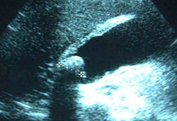
Use ultrasound as the first investigation to identify the presence of gallstones.
Request magnetic resonance cholangiopancreatography (MRCP) if ultrasound has not detected common bile duct stones but the bile duct is dilated and/or liver function test results are abnormal.[4]
The findings of acute cholecystitis on MRI are:[34]
Thickening of the gallbladder wall (≥4 mm)
Enlargement of the gallbladder (long axis ≥8 cm, short axis ≥4 cm)
Gallstones or retained debris
Fluid accumulation around the gallbladder
Linear shadows in the fatty tissue around the gallbladder.
Consider endoscopic ultrasound (EUS) if MRCP does not allow a diagnosis to be made.[4]
Practical tip
EUS is good at detecting distal common bile duct stones. If MRCP does not show a stone but the patient has deranged liver function tests, EUS is an excellent test but invasive; therefore, have a high index of suspicion before requesting this test.
Consider further investigations and appropriate management as required if conditions other than gallstone disease are suspected.[4]
Use CT for diagnosing emphysematous cholecystitis.[34]
Evidence: Comparison of imaging studies
Evidence shows that several imaging methods accurately rule out cholecystitis, although the diagnostic accuracy and costs of investigations vary.
Despite evidence that cholescintigraphy is more accurate than ultrasound and MR imaging at diagnosing acute cholecystitis, other factors such as availability, cost, and the ability to view an area outside of the biliary tract mean that ultrasound is generally the preferred initial test.
A systematic review, including 57 studies, sought to estimate the diagnostic accuracy of:[45]
Cholescintigraphy
Ultrasonography
MRI.
It found that the sensitivity of cholescintigraphy (96%, 95% CI 94% to 97%) was significantly higher than the sensitivity of ultrasonography (81%, 95% CI 75% to 87%) and magnetic resonance imaging (85%, 95% CI 66% to 95%) for diagnosing acute cholecystitis. There were no significant differences in specificity between cholescintigraphy (90%, 95% CI 86% to 93%), ultrasonography (83%, 95% CI 74% to 89%), and MR imaging (81%, 95% CI 69% to 90%).[45]
The 2018 Tokyo guidelines and the UK 2014 National Institute for Health and Care Excellence (NICE) guidelines both recommend ultrasound as a reasonable initial choice, based on issues such as low invasiveness, low risk, widespread availability, ease of use, and cost‐effectiveness.[4][34]
The NICE guideline recommends MRCP if abnormalities are present in the bile duct or liver function tests but ultrasound has not detected common bile duct stones.[4]
Two health economic studies found that MRCP appeared cost-effective compared with endoscopic retrograde cholangiopancreatography for diagnosing common bile duct stones.[4]
Note that in a patient with sepsis, use CT (or MRI) to identify the cause.
Full blood count
Look for significant deviations from the normal values.
A significantly raised or lowered white cell count can indicate an infection or inflammation.
C-reactive protein
Look for elevation, which may indicate infection or inflammation of the gallbladder.[27]
Bilirubin
Look for elevation, which may indicate acute focal cholestasis in adjacent liver tissue or be due to common bile duct stones.[4][27]
Liver function tests
Request liver function tests to indicate whether further imaging is required, such as magnetic resonance cholangiopancreatography.
May show elevated bilirubin, alkaline phosphatase, and gamma glutamyl transferase due to acute focal cholestasis in adjacent liver tissue or due to common bile duct stones.[4][27]
Alanine aminotransferase can also be elevated if a stone has passed down the common bile duct, or if there is focal inflammation of the liver parenchyma in severe cholecystitis.
Serum lipase or amylase
Identify or exclude the presence of acute pancreatitis.[27][33] Use serum lipase testing (if available) in preference to serum amylase.[33][46][47]
A result >3 times the upper limit of the normal range confirms the diagnosis of acute pancreatitis in a patient with acute upper abdominal pain.[33][48]
Serum lipase and amylase have similar sensitivity and specificity but lipase levels remain elevated for longer (up to 14 days after symptom onset versus 5 days for amylase), providing a higher likelihood of picking up the diagnosis in patients with a delayed presentation.[33][49][50]
Blood cultures and/or bile cultures
Request in patients with grade II (moderate) and III (severe) disease in order to identify an infection that may be the cause of sepsis.[51]
See Assessing severity below for guidance on how to define grade of cholecystitis.
Practical tip
There are no blood tests that will specifically confirm the diagnosis of cholecystitis, but they help to build the clinical picture of how unwell the patient is and can help to exclude other diagnoses.
Urgently refer or admit to hospital anyone with suspected acute cholecystitis.[27]
Sepsis and organ failure are of paramount importance in assessing severity.
During admission, assess severity based on the signs and symptoms of sepsis and the absence/presence of local complications or organ failure.
Assess the severity in order to determine the optimum treatment strategy.[52]
The Tokyo guidelines use a grading system of mild, moderate, and severe to classify severity. Treatment can be based on this classification.[34]
Severe (grade III) acute cholecystitis is associated with dysfunction of any one of the following organs/systems.[34]
Cardiovascular: hypotension requiring treatment with dopamine ≥5 micrograms/kg per minute, or any dose of noradrenaline (norepinephrine).
Neurological: decreased level of consciousness.
Respiratory: PaO 2/fraction of inspired oxygen (FiO 2) ratio <300.
Renal: oliguria, creatinine >2.0 mg/dL.
Hepatic: prothrombin time – international normalised ratio (PT‐INR) >1.5.
Haematological: platelet count <100,000/mm 3.
Moderate (grade II) acute cholecystitis is associated with any one of the following conditions.
Elevated white blood cell count (>18,000/mm 3).
Palpable tender mass in the right upper abdominal quadrant.
Duration of symptoms >72 hours.
Marked local inflammation (gangrenous cholecystitis, pericholecystic abscess, hepatic abscess, biliary peritonitis, emphysematous cholecystitis).
Mild (grade I) acute cholecystitis:[34]
Acute cholecystitis that does not meet the criteria of grade III or grade II acute cholecystitis. It can also be defined as acute cholecystitis in a healthy patient with no organ dysfunction and mild inflammatory changes in the gallbladder, making cholecystectomy a safe and low-risk operative procedure.
Use of this content is subject to our disclaimer