History and exam
Key diagnostic factors
common
presence of risk factors
Factors strongly associated with the risk of developing scleroderma include family history of the disease, and immune dysregulation (e.g., presence of antinuclear antibody [ANA]).
Raynaud's phenomenon
Must be differentiated from primary Raynaud's phenomenon, which is generally milder with no history of digital ulcerations and usually has a negative ANA.[18][19]
Secondary Raynaud's phenomenon is suggested by onset at over 30 years of age, intense asymmetric episodes associated with ischaemic ulcerations, other clinical features suggestive of connective tissue disease (e.g., inflammatory arthritis, abnormal pulmonary function tests [PFTs]), and evidence of micro-vascular disease on nail-fold capillaroscopy.[15][16][Figure caption and citation for the preceding image starts]: Hands demonstrating Raynaud's phenomenonFrom the collection of Maureen D. Mayes, University of Texas [Citation ends].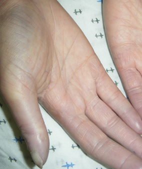
digital pits or ulcers
Ulcers occur in up to 50% of patients.
Located on distal digits or overlying bony prominences.
Can be painful, heal slowly, and lead to functional disability.
Ulcers on digital tips are thought to originate solely from ischaemia. [Figure caption and citation for the preceding image starts]: Digital tip infarctionFrom the collection of Maureen D. Mayes, University of Texas [Citation ends].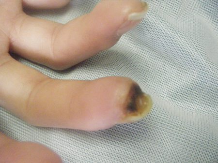 [Figure caption and citation for the preceding image starts]: Digital tip infarctionFrom the collection of Maureen D. Mayes, University of Texas [Citation ends].
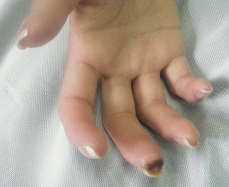
Digital pits with no ulceration may also be present. [Figure caption and citation for the preceding image starts]: Digital pits without ulcersFrom the collection of Maureen D. Mayes, University of Texas [Citation ends].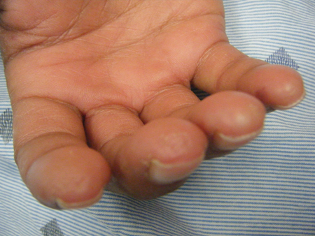
swelling of the hands and feet
May be an early manifestation of the condition, even before the onset of Raynaud's phenomenon.
Hand swelling usually associated with hand stiffness and most commonly worse in mornings.
Foot swelling prompts investigations into heart failure, renal impairment, or nephrotic syndrome.
skin thickening
Diffuse cutaneous disease is characterised by skin changes proximal to the elbows, and/or the anterior chest or abdomen.
An explosive onset with rapid progression after onset of Raynaud's phenomenon is characteristic of diffuse disease.
Limited cutaneous disease is defined by skin thickening distal to the elbows and knees.
Involvement of the face is noted in both diffuse and limited disease. This may cause a decrease in oral aperture.
loss of function of hands
Particularly the inability to get a tight grasp on objects.
Occurs as a result of the development of sclerodactyly.
sclerodactyly
Thickening of skin of fingers. [Figure caption and citation for the preceding image starts]: Fingers demonstrating sclerodactyly with finger curling, shiny skin at the fingers, and telangiectasiasFrom the collection of Maureen D. Mayes, University of Texas [Citation ends].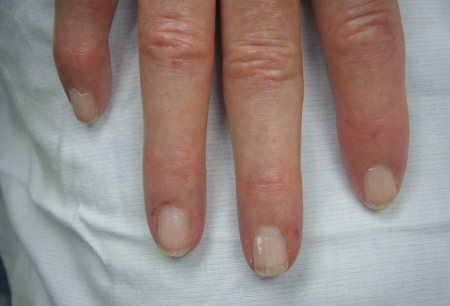 [Figure caption and citation for the preceding image starts]: Hand demonstrating sclerodactyly with finger curling, shiny skin at the fingers, and telangiectasiasFrom the collection of Maureen D. Mayes, University of Texas [Citation ends].
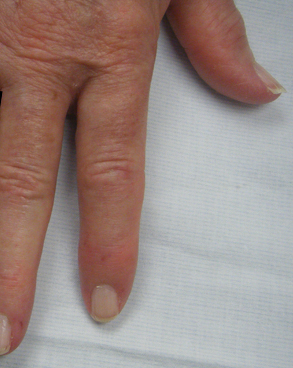 [Figure caption and citation for the preceding image starts]: Late-stage sclerodactyly with contractures due to severe skin tighteningFrom the collection of Maureen D. Mayes, University of Texas [Citation ends].
 [Figure caption and citation for the preceding image starts]: Late-stage sclerodactyly with contractures due to severe skin tighteningFrom the collection of Maureen D. Mayes, University of Texas [Citation ends].
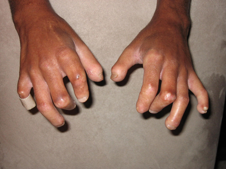
heartburn, reflux, and dysphagia
Heartburn and dysphagia are among the most common symptoms described in scleroderma.
Oesophageal dysmotility and incompetence of the lower oesophageal sphincter are found.
Treatment is necessary to prevent long-term consequences including oesophageal strictures, Barrett's metaplasia, and adenocarcinoma.[20][21]
bloating
Bloating is a common symptom due to decreased motility in the stomach and small bowel. Decreased small bowel motility can lead to bacterial overgrowth and malabsorptive diarrhoea, steatorrhoea, and weight loss.
faecal incontinence
Faecal incontinence may occur due to loss of tone of the anal sphincter.
arthralgias and myalgias
May be a presenting symptom, although non-specific.
abnormal nail-fold capillaroscopy
Indicative of changes on micro-vasculature that occur even early in disease.
Can also help in differentiating from primary Raynaud's phenomenon where capillaroscopy should be normal.
telangiectasia
Most commonly located on the fingers, palms, face, and mucous membranes. [Figure caption and citation for the preceding image starts]: Telangiectasias on the handFrom the collection of Maureen D. Mayes, University of Texas [Citation ends].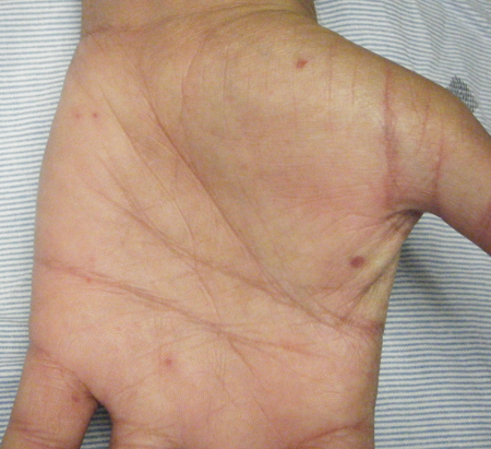 [Figure caption and citation for the preceding image starts]: Telangiectasias on the fingersFrom the collection of Maureen D. Mayes, University of Texas [Citation ends].
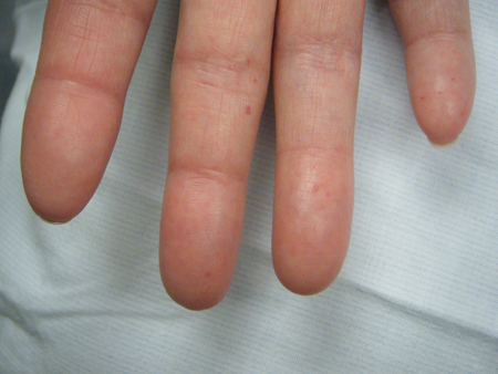
Can be numerous in the stomach, are referred to as 'watermelon stomach' (gastric antral vascular ectasia), and can cause chronic gastrointestinal bleeding and anaemia.
subcutaneous calcinosis
More common in long-standing disease.
Can present as small, localised, firm masses usually on fingers, forearms, or other pressure points.
Seen on plain x-rays.
Superficial lesions can open and become secondarily infected.
dyspnoea
Can be a sign of interstitial lung disease, pulmonary hypertension, chronic aspiration pneumonitis, or cardiomyopathy.
Yearly echocardiograms and complete PFTs (spirometry, lung volumes, and diffusing capacity) should be done for surveillance in scleroderma patients to screen for the development of some of these conditions.
Early detection is important, as treatment at this stage can be effective.
dry crackles at lung bases
Can be indicative of interstitial lung disease.
PFTs, chest x-ray, or high-definition CT should be performed if there is clinical suspicion for interstitial lung disease, particularly with restrictive pattern on PFTs.
Heart failure should be excluded if wet crackles are present.
uncommon
tendon friction rub
Palpable, and sometimes audible rub on movement of the ankle, wrist, knee, shoulder, or elbow, caused by irritation of the tendon sheath and/or adjacent tissues.
Usually found in diffuse disease.
A marker for aggressive disease with increased risk for internal organ involvement.[22]
abrupt onset moderate/marked hypertension
Can be presenting symptom of scleroderma in a small percentage of patients.
Is a sign of scleroderma renal crisis, which develops in 10% to 15% of patients, most commonly in the early stages of those with diffuse disease.
Is due to ischaemic activation of the renin-angiotensin system.
Scleroderma renal crisis is characterised by the onset of acute renal failure, abrupt onset of moderate/marked hypertension, a urinary sediment that is frequently normal or reveals only mild proteinuria with few cells or casts, and a micro-angiopathic haemolytic anaemia.
Other findings of hypertensive emergency may be found (e.g., papilloedema, headache, confusion, decreased urine output, chest pain).
Other diagnostic factors
common
fatigue
May be an early manifestation of the condition and may continue through the disease course.
May be a subtle symptom of interstitial lung disease, pulmonary artery hypertension, or anaemia from gastrointestinal blood loss.
dry cough
Can be indicative of lung involvement by either interstitial lung disease or chronic aspiration.
decreased exercise tolerance
Can be indicative of cardiomyopathy or lung involvement by interstitial lung disease, pulmonary hypertension, or chronic aspiration.
Reduced level of physical fitness should also be considered.
uncommon
weight loss
May be an early sign and the cause of the initial presentation.
inflammatory arthritis
The presence of inflammatory arthritis may be suggestive of an overlap or mixed connective tissue disease.
May be rheumatoid-like with true erosions on x-ray.
proximal muscular weakness (inflammatory myositis)
Inflammatory myositis may be seen in a subset of patients with scleroderma.
It is differentiated from scleroderma myopathy (another type of muscular condition associated with scleroderma) by the presence of weakness (usually in the proximal muscles) and typical electromyogram/nerve conduction and muscle biopsy findings.
synovitis
Can be difficult to identify as independent from the diffuse hand swelling and overlying thickened skin.
Joint tenderness on compression or squeezing should be present.
Inflammatory markers (erythrocyte sedimentation rate, C-reactive protein) may be elevated.
increased accentuation of the pulmonic component of S2 heart sound
If present is suggestive of pulmonary hypertension.
signs of anaemia
Pallor, fatigue, and cardiac manifestations (tachycardia, heart murmurs, or cardiac enlargement) may be present. Commonly from chronic gastrointestinal (GI) blood loss from gastric antral vascular ectasia.
This can be a major source of morbidity in scleroderma patients.
Should be further investigated by upper GI endoscopy.
Risk factors
strong
family history of scleroderma
A genetic predisposition is suggested by a study showing the increased prevalence of scleroderma among first-degree relatives of affected probands.[10]
Scleroderma is associated with human leukocyte antigen class II alleles.[13] It is also associated with polymorphisms of multiple other genes, most of which affect immune function.[14]
immune dysregulation (e.g., positive ANA)
Auto-antibodies are associated with distinct clinical phenotypes of scleroderma.
About 90% of patients have a positive ANA. Among this group, there are subsets of mutually exclusive auto-antibodies that are associated with distinct clinical phenotypes.
Anti-topoisomerase I (anti-Scl 70) antibody is seen in about 20% of cases and is associated with an increased risk of interstitial lung disease and with diffuse skin involvement.
Anti-RNA polymerase III antibody (about 20% of cases) is associated with renal crisis.
Anti-centromere antibodies (20% to 25% of cases) are associated with limited skin involvement and a better overall prognosis.
Other scleroderma-specific antibodies, such as anti-fibrillarin (also known as U3RNP or anti Th-To), are not readily available commercially, and together represent only about 5% to 10% of all SSc patients.
The remaining 35% to 40% of patients with scleroderma do not have an as-yet-identified scleroderma-specific auto-antibody.
weak
exposure to environmental substances and toxins (e.g., silica dust or solvents)
Several factors have been postulated to have an association with scleroderma, including exposure to silica dust and multiple solvents, but no clear link has been identified.[12]
Use of this content is subject to our disclaimer